Copyright
©The Author(s) 2023.
World J Clin Cases. Jul 26, 2023; 11(21): 5035-5046
Published online Jul 26, 2023. doi: 10.12998/wjcc.v11.i21.5035
Published online Jul 26, 2023. doi: 10.12998/wjcc.v11.i21.5035
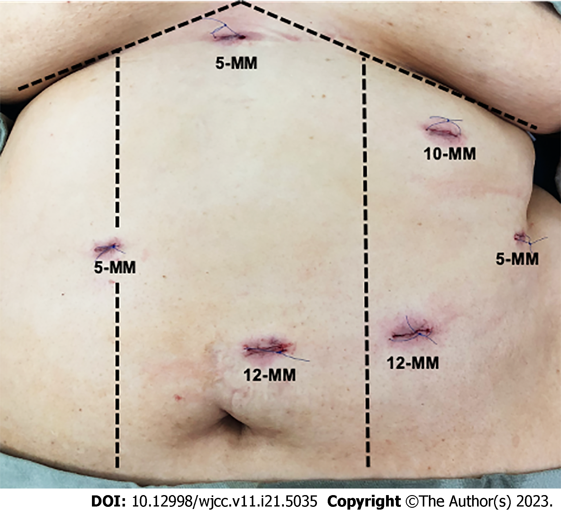
Figure 1 Positioning of trocars for laparoscopic single-anastomosis duodeno-ileal bypass with sleeve gastrectomy.
Figure 2 Sequence of single-anastomosis duodenum ileal bypass with sleeve gastrectomy.
A: Dissection of duodenum posterior to gastroduodenal artery; B and C: Forty-five mm stapled line of the duodenum, leaving 5 cm of the duodenum proximal to the pylorus; D: End-to-side duodenal ileostomy in two planes and methylene blue test without leaks; C: Common bile duct; D: Duodenum; AA: Afferent loop; AE: Efferent loop; P: Pylorus; T: Portal triad; DP: Proximal duodenum; DD: Distal duodenum; S: stomach.
Figure 3 Frequency distribution of laboratory results taken before surgery (T1), at 6 mo (T2), and at one year (T3) of follow-up.
A: The median hemoglobin was 14.35 mg/dL [interquartile range (IQR) 1.82] at T1, 13.47 mg/dL (IQR 2.6) at T2, and 13.50 mg/dL (1.37) at T3; B: The median hematocrit was 44.36% (IQR 5.17) at T1, 30.27% (IQR 6.5) at T2, and 40.22% (3.33) at T3; C: Median glycated hemoglobin was 5.81% (IQR 0.75) at T1, 5.1% (IQR 0.48) at T2, and 13.50% (IQR 0.51) at T3; D: Median glucose levels were 97.54 mg/dL (IQR 21.07) at T1, 81.7 mg/dl (IQR 10.99) at T2, and 79 mg/dL (IQR 10.9) at T3; E: Median TSH levels were 2.8 mU/L at T1, 2.10 mU/L at T2, and 2.46mU/L at T3 ; F: Cholesterol at T1 of 179.80 mg/dL (IQR 48.9) dropped at T2 to 143.38 mg/dL (IQR 46) and was maintained at T3 at 143.13 mg/dL (IQR 26); G: High-density lipoprotein levels (HDL) went from 44.1 mg/dL (IQR 10.9) at T1 to 41.35 mg/dL (IQR 12.45) at T2 and 46.5 mg/dL (IQR 13) at T3; H: LDL levels went from 108 mg/dL (IQR 39.6) at T1 to 89.4 mg/dL (IQR 36.31) at T2 and 84.37 mg/dL (IQR 37.67) at T3; I: Triglyceride levels went from 110.8 mg/dL (IQR 64.75) at T1 to 90 mg/dL (IQR 46.09) at T2 and 75.5 mg/dL (IQR 25.8) at T3. T1: Preoperative, T2: 6 mo, T3: 1 year. aP < 0.05, bP < 0.01, cP < 0.01, dP < 0.00001. NS: Not significant.
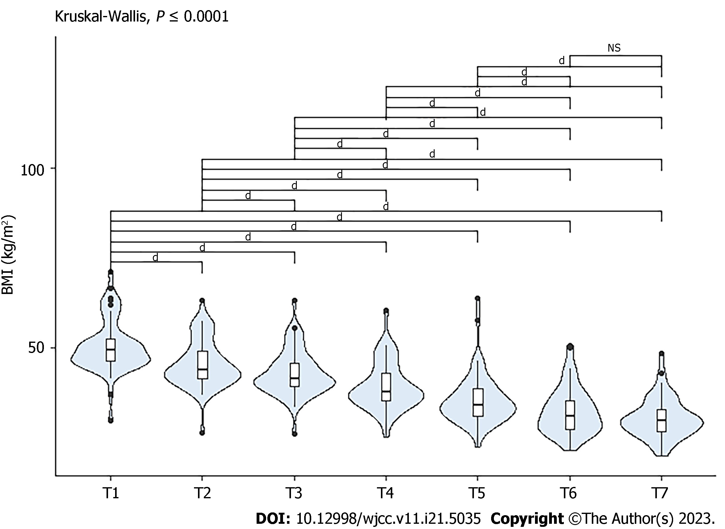
Figure 4 Frequency distribution of body mass index of patients during follow-up.
T1: Pre-surgical, median body mass index (BMI) 49.15 kg/m2 [interquartile range (IQR) 5.98]. Postoperative: T2: 2 wk, median BMI 43.73 kg/m2 (IQR 7.49); T3: 1 mo, median BMI 41.22 kg/m2 (IQR 6.39); T4: 3 mo, median BMI 37.52 kg/m2 (IQR 7.56); T5: 6 mo, median BMI 33.91 kg/m2 (IQR 7.71); T6: 1 year, median BMI 30.99 kg/m2 (IQR 8.02); T7: 2 years, median BMI 29.61 kg/m2 (IQR 6.04). dP < 0.00001. NS: Not significant.
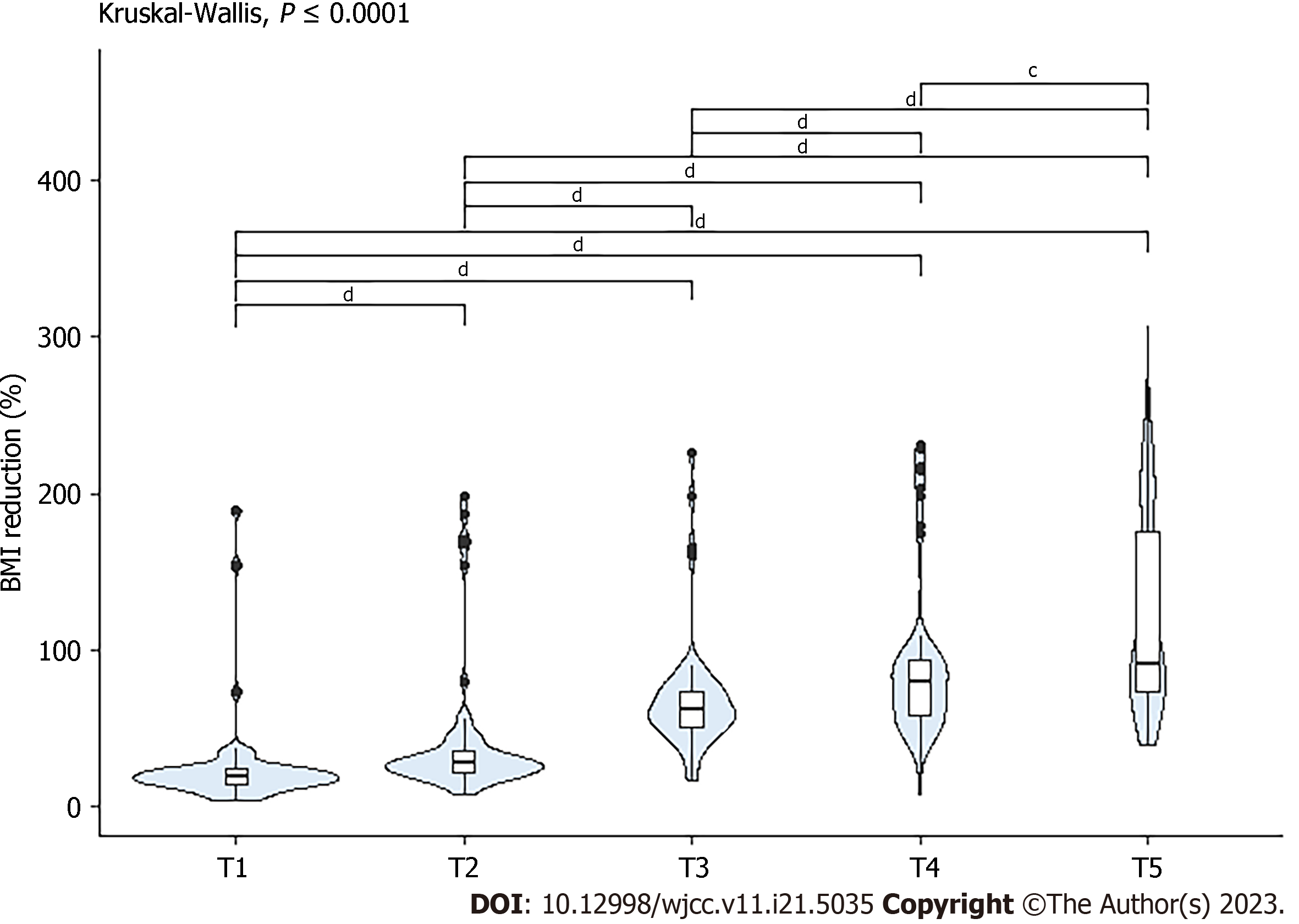
Figure 5 Frequency distribution of percentage reduction in body mass index of patients during follow-up.
Postoperative: T1: 2 wk, median body mass index (BMI) reduction 19.53% [interquartile range (IQR) 9.57]; T2: 3 mo, median BMI reduction 27.58% (IQR 14.46); T3: 6 mo, median BMI reduction 61.71% (IQR 23.78); T4: 1 year, median BMI reduction 79.84% (IQR 35.76); T5: 2 years, median BMI reduction 91.27% (IQR 102.23). cP < 0.01, dP < 0.00001.
- Citation: Ospina Jaramillo A, Riscanevo Bobadilla AC, Espinosa MO, Valencia A, Jiménez H, Montilla Velásquez MDP, Bastidas M. Clinical outcomes and complications of single anastomosis duodenal-ileal bypass with sleeve gastrectomy: A 2-year follow-up study in Bogotá, Colombia. World J Clin Cases 2023; 11(21): 5035-5046
- URL: https://www.wjgnet.com/2307-8960/full/v11/i21/5035.htm
- DOI: https://dx.doi.org/10.12998/wjcc.v11.i21.5035


