Copyright
©The Author(s) 2023.
World J Clin Cases. Jul 16, 2023; 11(20): 4944-4955
Published online Jul 16, 2023. doi: 10.12998/wjcc.v11.i20.4944
Published online Jul 16, 2023. doi: 10.12998/wjcc.v11.i20.4944
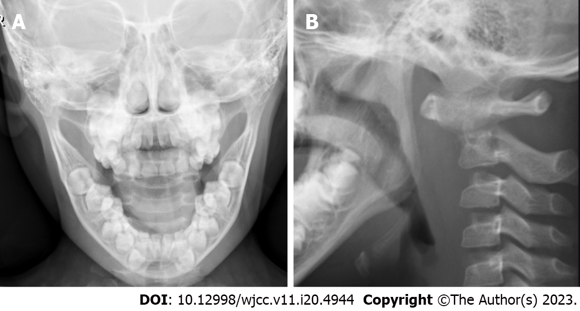
Figure 1 Preoperative open-mouth and lateral X-rays of the atlantoaxial joint.
Axial bone structural disorder, localized loss of bone density, and narrowing of the atlantoaxial joint space were all shown on preoperative X-ray films. A: Open-mouth view; B: Lateral view.
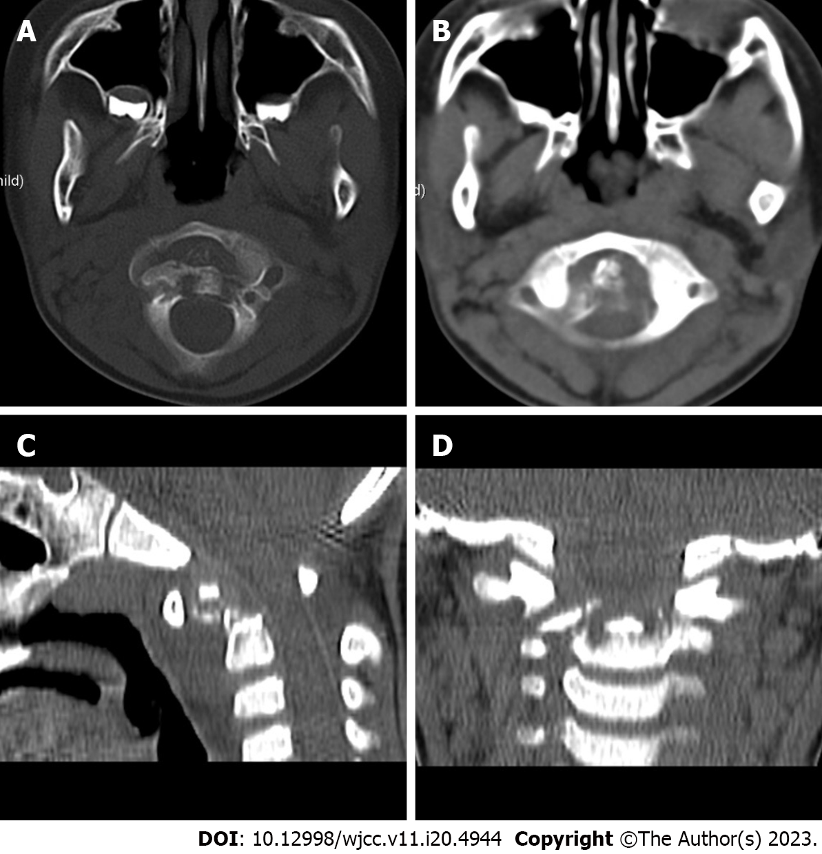
Figure 2 Preoperative computed tomography of the atlantoaxial joint.
Atlantoaxial subluxation and bone destruction at the top edge of the axial vertebral body were the preoperative computed tomography findings. A: Bone window; B: Soft tissue window; C: Sagittal reconstruction; D: Coronal reconstruction.
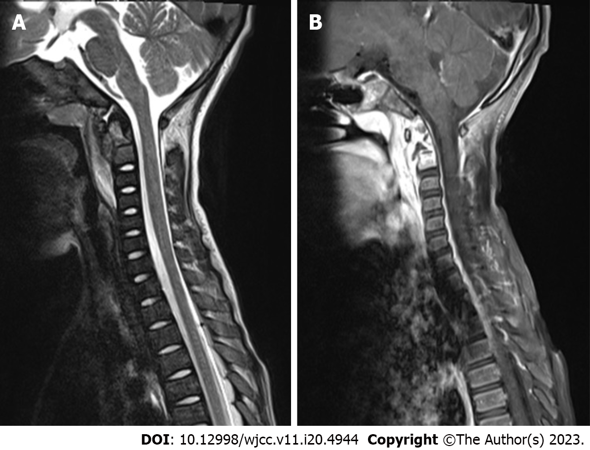
Figure 3 Preoperative magnetic resonance imaging of the cervical spine.
Cervical spine magnetic resonance imaging with contrast enhancement revealed odontoid destruction and abnormal signals in the retropharyngeal and prevertebral spaces. A: T2-weighted sagittal imaging of cervical vertebrae; B: Contrast-enhanced T1-weighted sagittal imaging of cervical vertebrae.
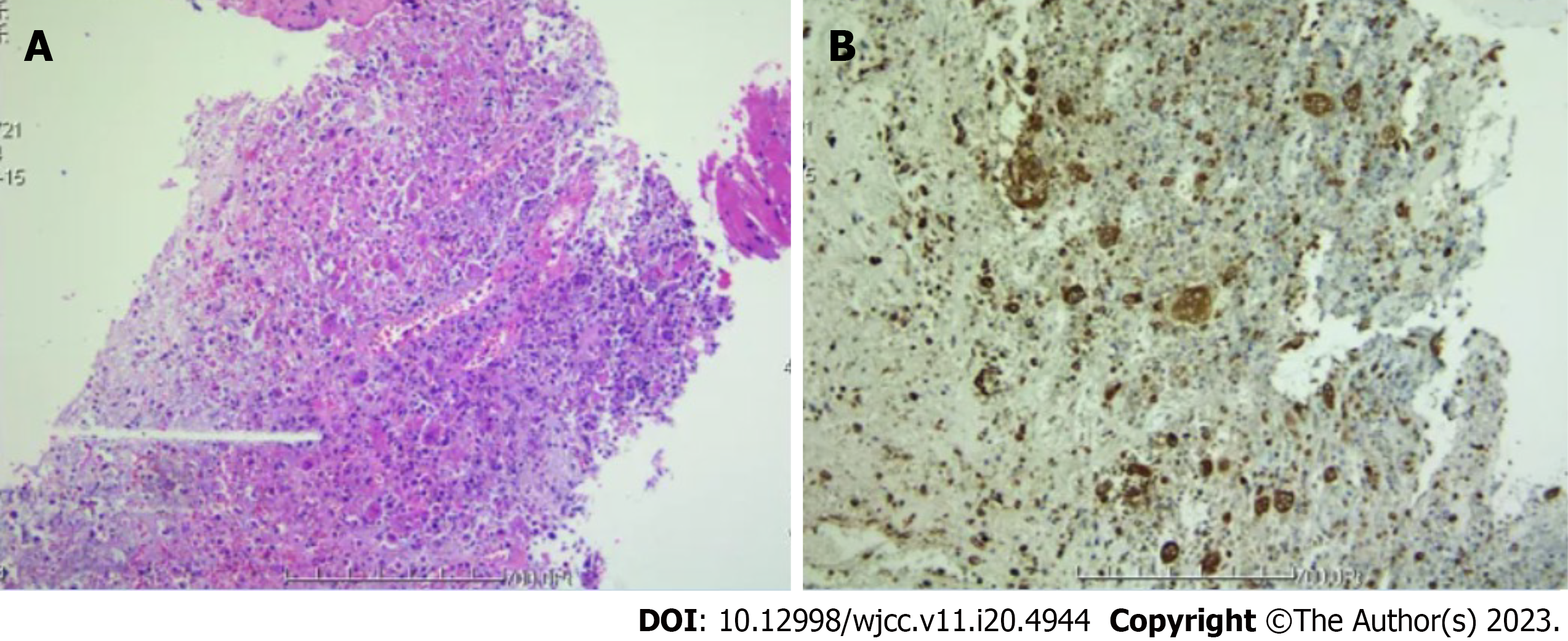
Figure 4 Preoperative pathological examination of tissue specimens.
Transoral endoscopic biopsy of the posterior pharyngeal wall lesion indicated an axial eosinophilic granuloma. A: Hematoxylin and eosin staining; B: Immunohistochemistry.
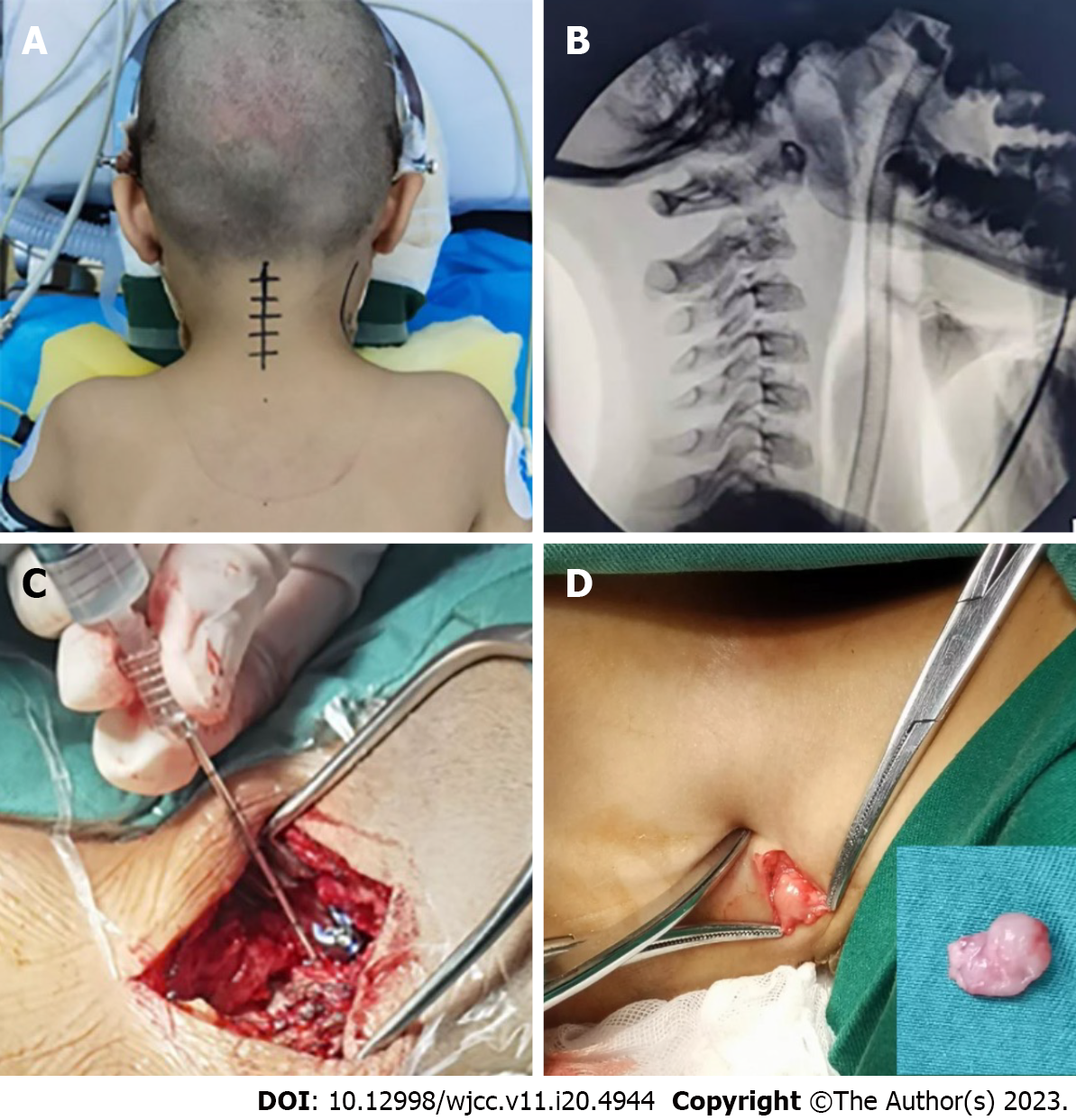
Figure 5 Hyperextension positioning with skull traction.
Figure 6 The pictures during the operation.
A: The patient lay prone on the plaster bed for reduction; B: Intraoperative view from the atlantoaxial side; C: Through the bilateral C2 pedicle screw channels, 80 mg of methylprednisolone was injected into the C2 vertebral lesions; D: The boy's intraoperative cervical lymph nodes.
Figure 7 Postoperative open-mouth and lateral X-rays of the atlantoaxial joint.
Atlantoaxial joint dislocation was successfully corrected by the posterior atlantoaxial pedicle screw, according to the postoperative X-ray. A: Open-mouth view; B: Lateral view.
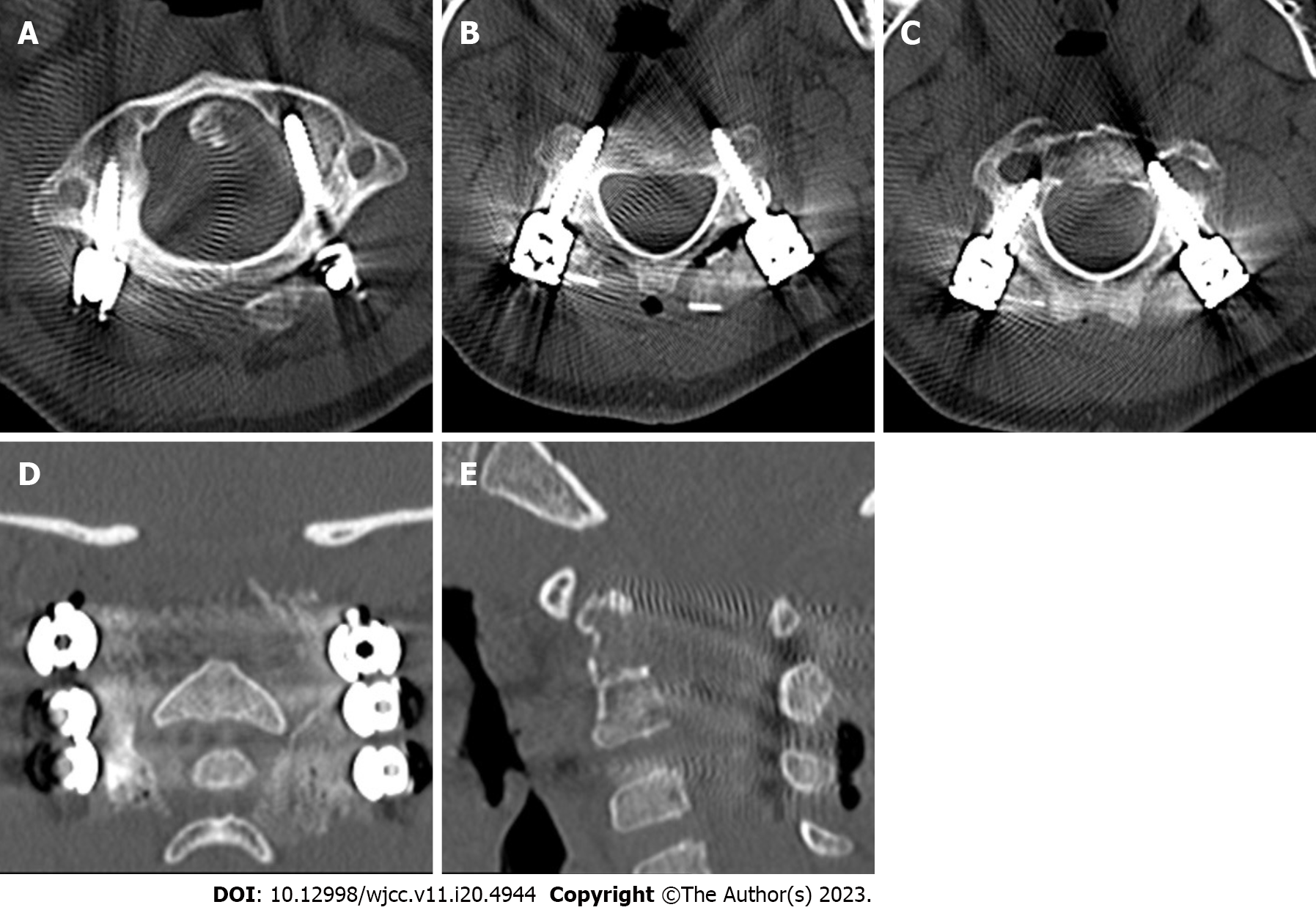
Figure 8 Postoperative computed tomography of the atlantoaxial joint.
The atlantoaxial joint's dislocation was corrected, the screw position was good, and the bone graft had been successfully imbedded, according to a postoperative computed tomography scan. A-C: Bone window of the atlas vertebra, axial section; D: Coronal reconstruction; E: Sagittal reconstruction.
Figure 9 Postoperative magnetic resonance imaging of the atlantoaxial joint.
There was less atlantoaxial joint dislocation, the screw position was good, there was no spinal canal stenosis, the contour of the spinal cord was normal, the signal was uniform, and no abnormal signal was identified in the spinal canal. A: T2-weighted non-fat-saturation sequence; B: T2-weighted fat suppression sequence; C: T1-weighted plain scan.
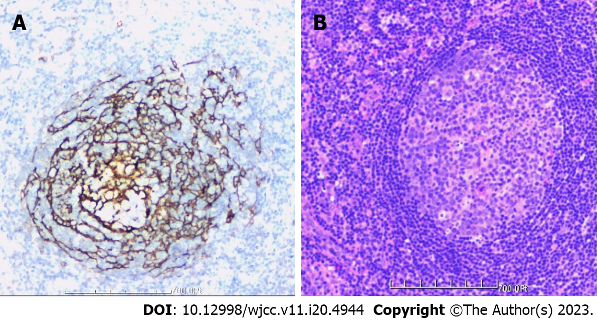
Figure 10 Postoperative pathological examination of tissue specimens.
The tissue was primarily hyperplastic and degenerative fibrous tissue and degenerative bone tissue, with flaky distribution, partial necrosis of bone tissue, interstitial vascular hyperplasia and dilatation with inflammatory cell infiltration, patchy areas of necrosis (pink), and a significant amount of multinucleated giant-cell infiltration. Plasma cell CD38 (focus +), CD138, lymphocyte LCA (+), Ki-67 approximately 10% graded CK (-), EMA (-), Smur100 (+), CD68 (histiocyte +), CD1a (histiocyte +), SMA (vascular +), and CD34 (vascular +) were all detected in the tissue by immunohistochemistry. The diagnosis of EG was supported by histology, immunohistochemical staining, and other factors. A: Immunohistochemistry; B: Hematoxylin and eosin staining.
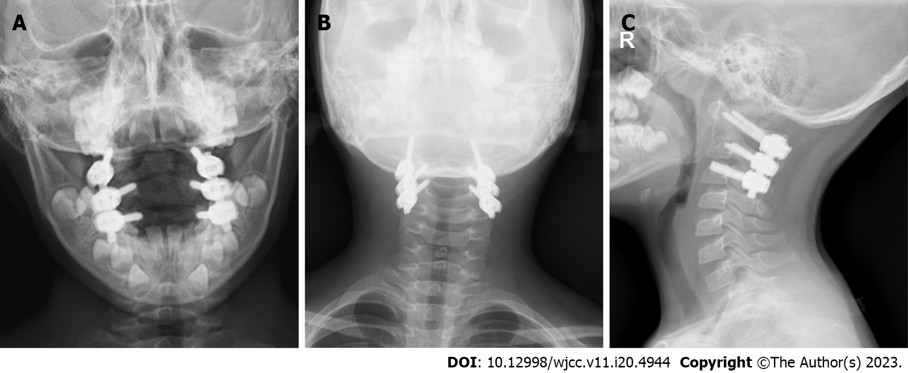
Figure 11 Postoperative X-ray of cervical spine one year later.
The atlantoaxial dislocation had been repaired and was in a satisfactory fixed position one year after the procedure, according to an X-ray. A: Open-mouth view; B: Anterior posterior view; C: Lateral view.
Figure 12 Postoperative computed tomography of cervical spine one year later.
The internal fixation was in a favorable position, the iliac bone graft was well fused, and the atlantoaxial dislocation had healed one year after the operation, according to the computed tomography (CT) scan. A and B: CT bone window of atlas axis; C: CT sagittal reconstruction of atlas axis; D: CT coronal reconstruction of atlas axis.
Figure 13 Postoperative magnetic resonance imaging of cervical spine one year later.
One year following the surgery, magnetic resonance imaging (MRI) revealed that the axis lesion had greatly shrunk and that the atlantoaxial joint was normal. A: T2-weighted MRI scan, sagittal view; B: T1-weighted MRI scan, sagittal view.
- Citation: Tu CQ, Chen ZD, Yao XT, Jiang YJ, Zhang BF, Lin B. Posterior pedicle screw fixation combined with local steroid injections for treating axial eosinophilic granulomas and atlantoaxial dislocation: A case report. World J Clin Cases 2023; 11(20): 4944-4955
- URL: https://www.wjgnet.com/2307-8960/full/v11/i20/4944.htm
- DOI: https://dx.doi.org/10.12998/wjcc.v11.i20.4944


