Copyright
©The Author(s) 2023.
World J Clin Cases. Jul 16, 2023; 11(20): 4897-4902
Published online Jul 16, 2023. doi: 10.12998/wjcc.v11.i20.4897
Published online Jul 16, 2023. doi: 10.12998/wjcc.v11.i20.4897
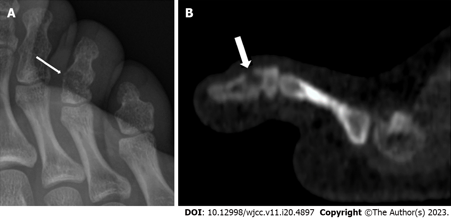
Figure 1 Imaging.
A: A plain radiograph performed after 2 mo of follow-up revealed an ill-defined lytic lesion of the fourth toe’s fused distal phalanx (white arrow); B: A computed tomography scan revealed an eccentrically-located lytic focus of the phalangeal metaphysis that was eroding the dorsal cortex (white arrow).
- Citation: Vazquez O, De Marco G, Gavira N, Habre C, Bartucz M, Steiger CN, Dayer R, Ceroni D. Subacute osteomyelitis due to Staphylococcus caprae in a teenager: A case report and review of the literature. World J Clin Cases 2023; 11(20): 4897-4902
- URL: https://www.wjgnet.com/2307-8960/full/v11/i20/4897.htm
- DOI: https://dx.doi.org/10.12998/wjcc.v11.i20.4897


