Copyright
©The Author(s) 2023.
World J Clin Cases. Jul 16, 2023; 11(20): 4890-4896
Published online Jul 16, 2023. doi: 10.12998/wjcc.v11.i20.4890
Published online Jul 16, 2023. doi: 10.12998/wjcc.v11.i20.4890
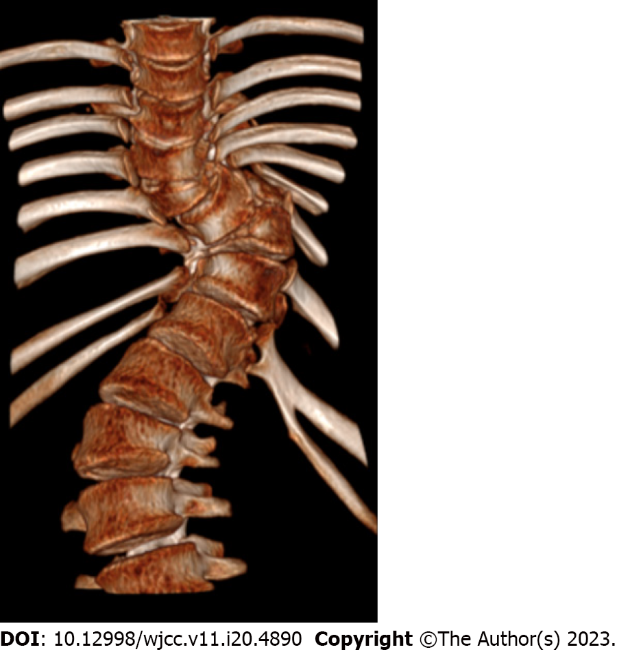
Figure 1 Three-dimensional computed tomography reconstruction of the preoperative spine.
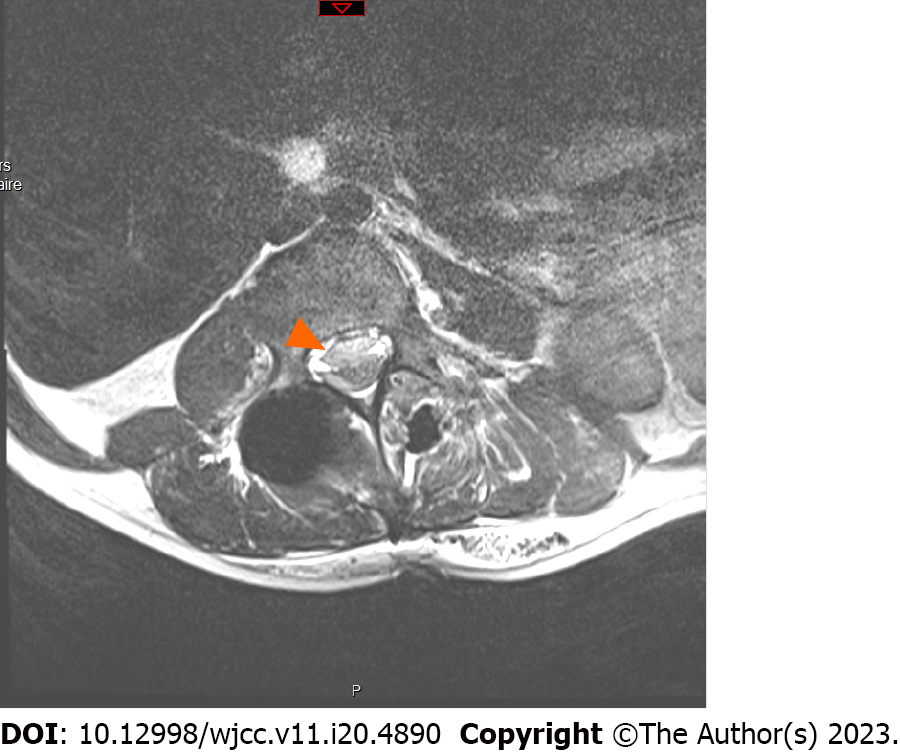
Figure 2 Axial view of L3 in T2-weighted magnetic resonance imaging.
The orange arrow shows an anterior haematoma displacing the spinal cord and nerve roots posteriorly.
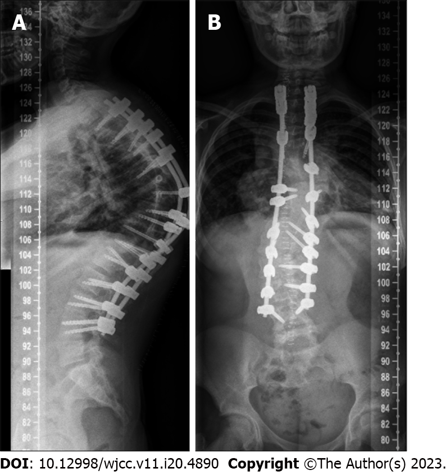
Figure 3 Postoperative X-rays.
A: Lateral view; B: Posteroanterior view.
- Citation: Michon du Marais G, Tabard-Fougère A, Dayer R. Acute spinal subdural haematoma complicating a posterior spinal instrumented fusion for congenital scoliosis: A case report. World J Clin Cases 2023; 11(20): 4890-4896
- URL: https://www.wjgnet.com/2307-8960/full/v11/i20/4890.htm
- DOI: https://dx.doi.org/10.12998/wjcc.v11.i20.4890


