Copyright
©The Author(s) 2023.
World J Clin Cases. Jul 16, 2023; 11(20): 4734-4739
Published online Jul 16, 2023. doi: 10.12998/wjcc.v11.i20.4734
Published online Jul 16, 2023. doi: 10.12998/wjcc.v11.i20.4734
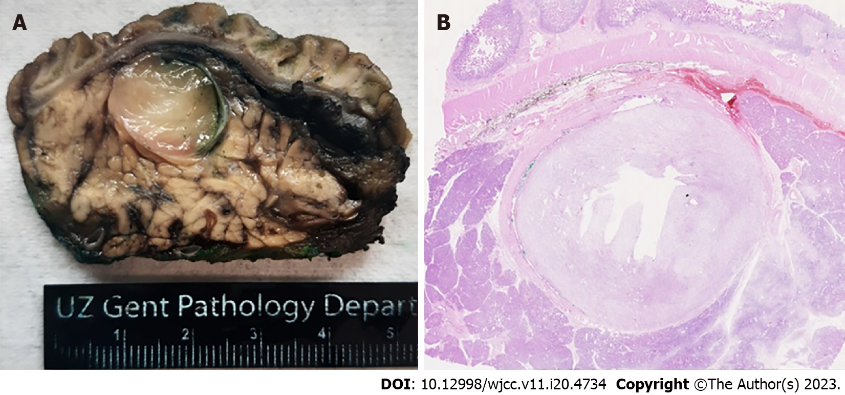
Figure 1 Inflammatory myofibroblastic tumor of the common bile duct.
A: Macroscopic picture showing a gelatinous lesion in the lumen of the intrapancreatic part of the common bile duct; B: Microscopic examination of the same lesion confirming its myxoid nature (hematoxylin and eosin, original magnification 10x).
Figure 2 Histological and immunohistochemical characteristics.
A: At high magnification this inflammatory myofibroblastic tumor is composed of plump spindle cells with a vesicular nucleus and pale cytoplasm. The stroma is myxoid with an inflammatory infiltrate composed of lymphocytes, plasma cells, macrophages and scarce eosinophils (hematoxylin and eosin, original magnification, 100x); B: The lesion demonstrates obvious ALK positivity in the spindle cells (original magnification 500x).
- Citation: Cordier F, Hoorens A, Ferdinande L, Van Dorpe J, Creytens D. Inflammatory myofibroblastic tumor of the distal common bile duct: Literature review with focus on pathological examination. World J Clin Cases 2023; 11(20): 4734-4739
- URL: https://www.wjgnet.com/2307-8960/full/v11/i20/4734.htm
- DOI: https://dx.doi.org/10.12998/wjcc.v11.i20.4734


