Copyright
©The Author(s) 2023.
World J Clin Cases. Jan 16, 2023; 11(2): 456-463
Published online Jan 16, 2023. doi: 10.12998/wjcc.v11.i2.456
Published online Jan 16, 2023. doi: 10.12998/wjcc.v11.i2.456
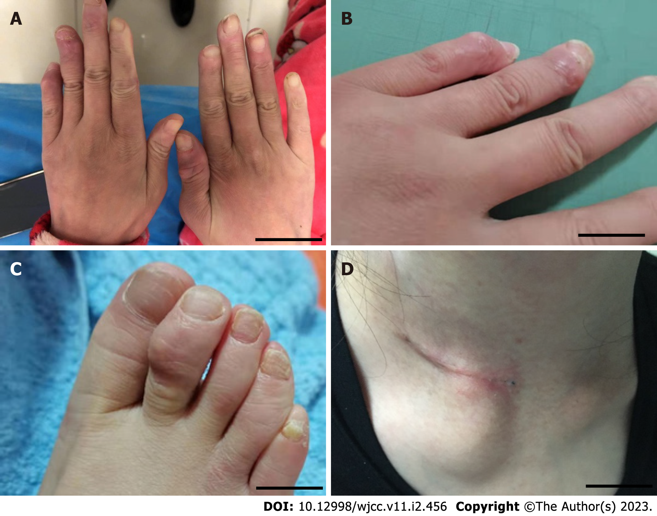
Figure 1 The patient was admitted to the hospital with new skin nodules on the distal interphalangeal of the hands and feet and a mass over the right sternoclavicular joint.
A and B: Papulonodular lesions over the interphalangeal joint of the both hands; C: Feet; D: Right sternoclavicular joint. Scale bar = 50 μm.
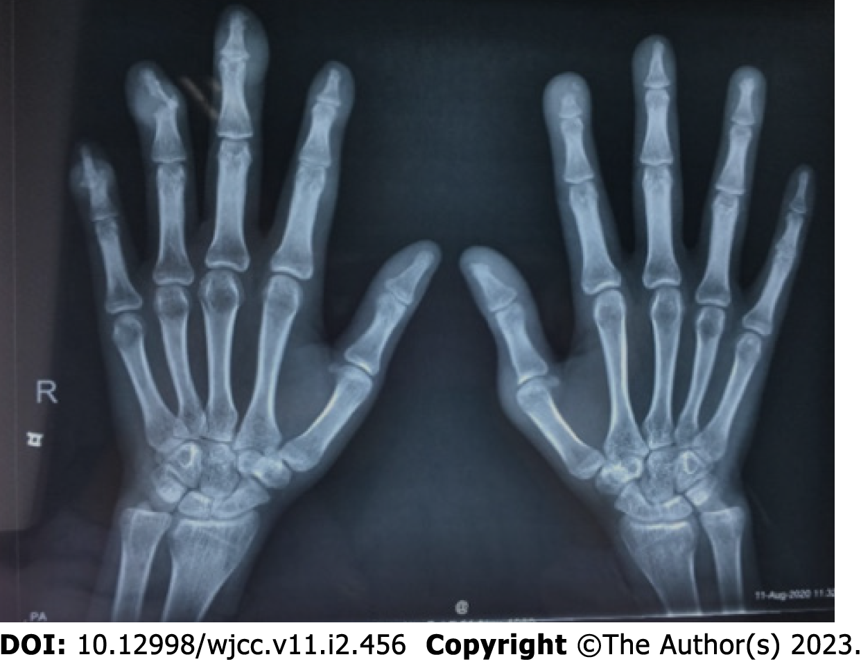
Figure 2 X-ray of both hands showed bone erosion at the distal interphalangeal joints with appearance of a pencil-in-cup deformity and articular soft tissue swelling.
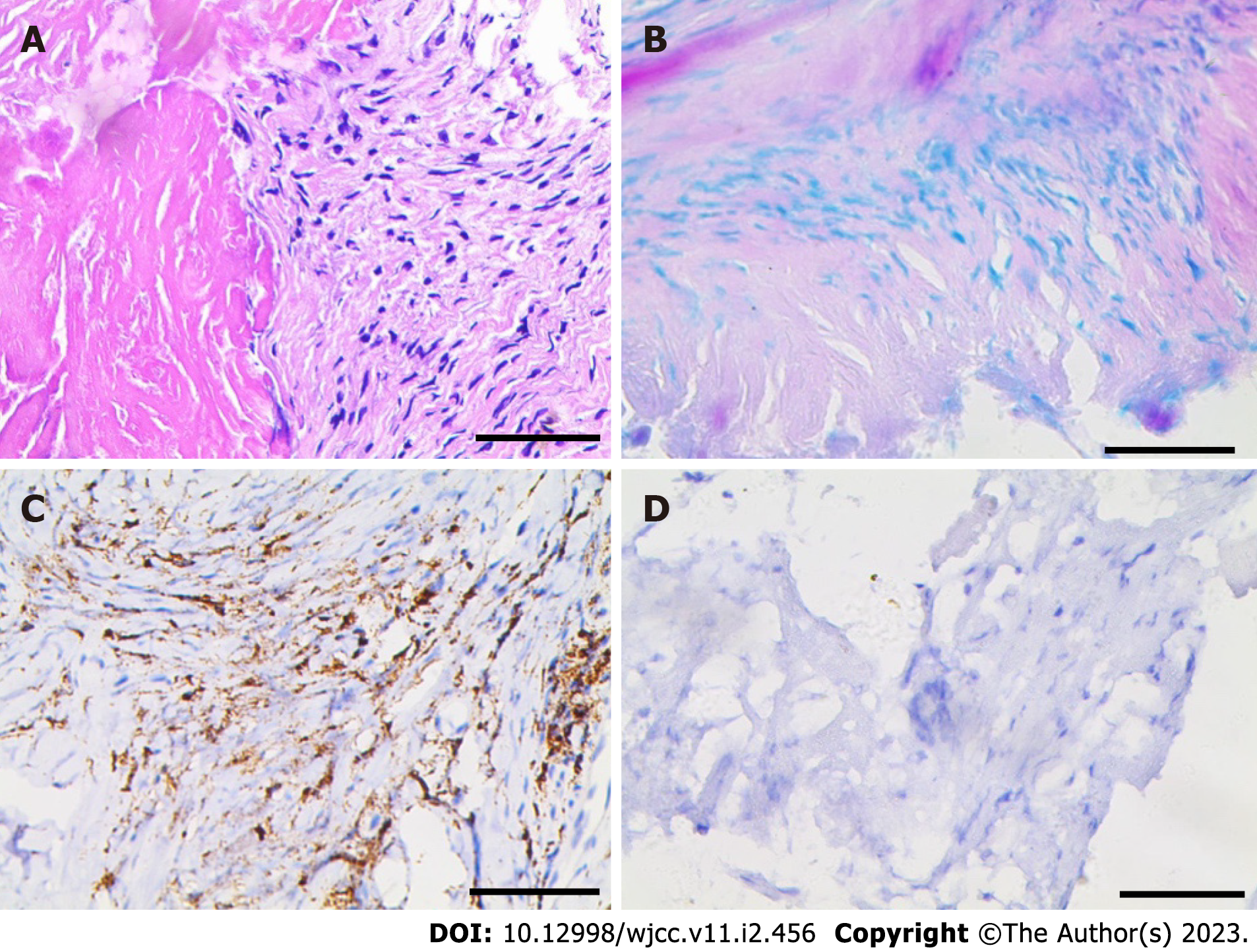
Figure 3 Immunohistochemical staining (with 40 × magnification).
A: Hematoxylin-eosin stain; B: Acid-fact stain; C: Positive for CD68; D: Negative for S100). Scale bar = 50 μm.
- Citation: Liu PP, Shuai ZW, Lian L, Wang K. Systemic lupus erythematosus with multicentric reticulohistiocytosis: A case report. World J Clin Cases 2023; 11(2): 456-463
- URL: https://www.wjgnet.com/2307-8960/full/v11/i2/456.htm
- DOI: https://dx.doi.org/10.12998/wjcc.v11.i2.456


