Copyright
©The Author(s) 2023.
World J Clin Cases. Jan 16, 2023; 11(2): 426-433
Published online Jan 16, 2023. doi: 10.12998/wjcc.v11.i2.426
Published online Jan 16, 2023. doi: 10.12998/wjcc.v11.i2.426
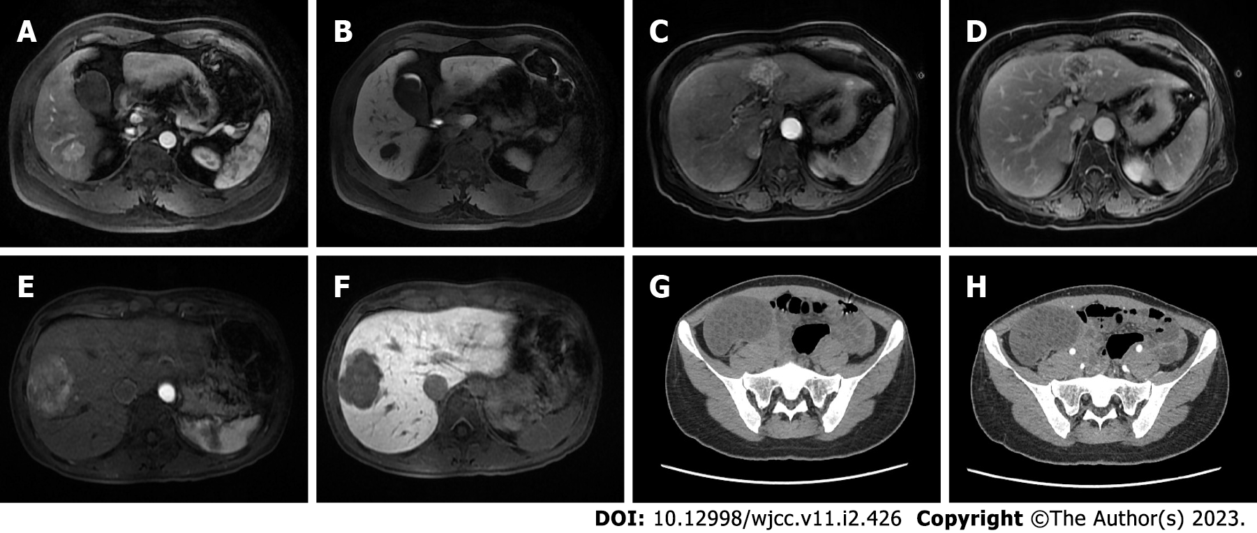
Figure 1 Three cases misdiagnosed as hepatocellular carcinoma based on imaging findings.
A and B: Case 1: Magnetic resonance imaging (MRI) of hepatic perivascular epithelioid cell neoplasms (PEComa) in a 37-year-old man: the lesion located in S6 showed distinct enhancement during the hepatic arterial phase (A), rim enhancement on the delayed phase (B); C and D: Case 2: MRI of hepatic PEComa in a 70-year-old woman, the lesion displayed obvious heterogeneous enhancement in the arterial phase (C), that was significantly weakened in the venous phase (D); E and F: Case 3: MRI of hepatic PEComa in a 30-year-old woman accompanied by an ovarian mature cystic teratoma: the lesions showed asymmetrical enhancement in arterial phase images in enhanced scanning (E), lesion enhancement was significantly weakened in the portal phase (F); G and H: preoperative plain abdominal computed tomography showed a right ovarian lesion with multiple cystic low-density shadows scattered throughout (G), the lesion was not strengthened after undergoing contrast-enhanced computed tomography (H).
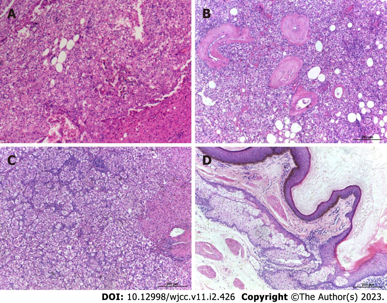
Figure 2 Results of hematoxylin-eosin staining.
A: Hematoxylin-eosin (HE) staining of the liver in case 1, a 37-year-old man; B: HE staining of the liver in case 2, a 70-year-old woman; C: HE staining of the liver in case 3, a 30-year-old woman; D: HE staining of the ovarian mature cystic teratoma in case 3, a 30-year-old woman.
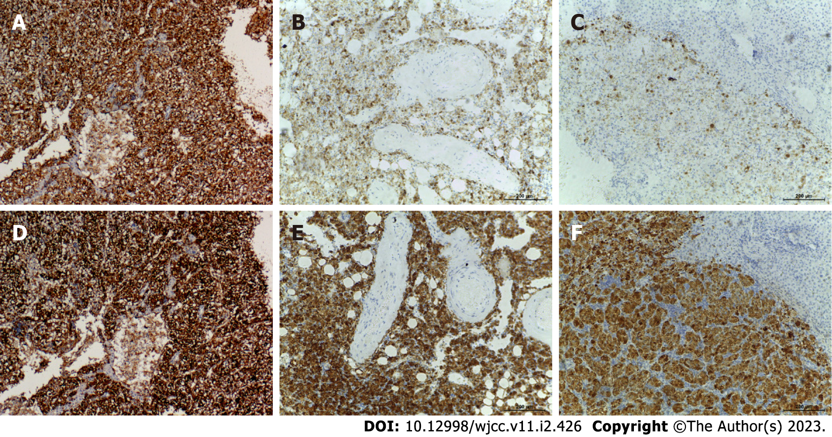
Figure 3 Immunohistochemical staining results indicated that all three patients had hepatic perivascular epithelioid cell neoplasm.
A: Positive staining for the biomarker human melanoma black 45 (HMB45) of case 1; B: Positive staining for HMB45 of case 2; C: Positive staining for HMB45 of case 3; D: Positive straining for the biomarker Melan A of case 1; E: Positive straining for Melan A of case 2; F: Positive straining for Melan A of case 3.
- Citation: Kou YQ, Yang YP, Ye WX, Yuan WN, Du SS, Nie B. Perivascular epithelioid cell tumors of the liver misdiagnosed as hepatocellular carcinoma: Three case reports. World J Clin Cases 2023; 11(2): 426-433
- URL: https://www.wjgnet.com/2307-8960/full/v11/i2/426.htm
- DOI: https://dx.doi.org/10.12998/wjcc.v11.i2.426


