Copyright
©The Author(s) 2023.
World J Clin Cases. Jan 16, 2023; 11(2): 408-416
Published online Jan 16, 2023. doi: 10.12998/wjcc.v11.i2.408
Published online Jan 16, 2023. doi: 10.12998/wjcc.v11.i2.408
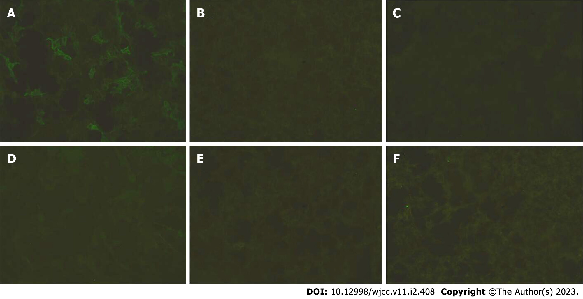
Figure 1 LGI-1(A), CASPR2(B), NMDAR(C), GAD65(D), GABA(E), AMPA1(F) antibodies in CSF validated by cell-assay of transfected cells.
LGI1-IgG in CSF (1:3.2) were positive but others (CASPR2, NMDAR, GAD65, GABA, AMPA1) were negative.
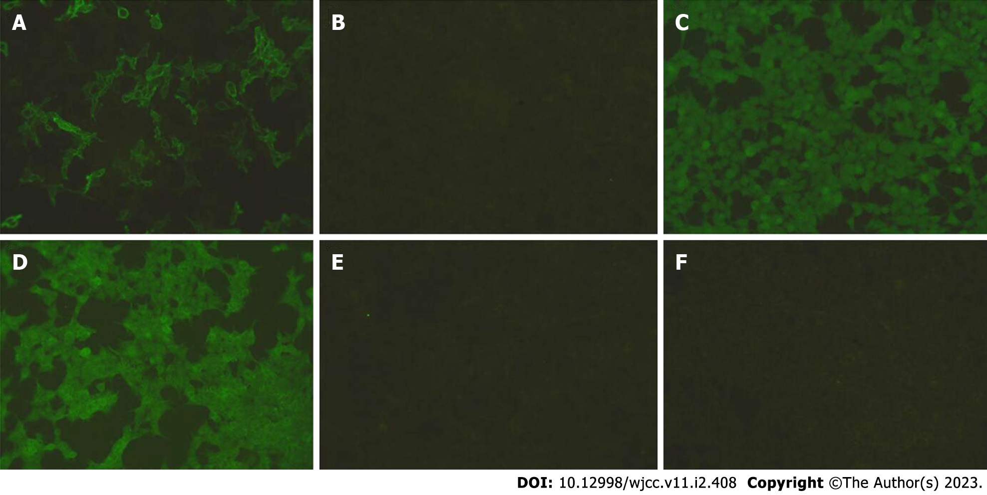
Figure 2 LGI-1(A), CASPR2(B), NMDAR(C), GAD65(D), GABA(E), AMPA1(F) antibodies in serum validated by cell-assay of transfected cells.
LGI1-IgG in serum (1:100) were positive but others (CASPR2, NMDAR, GAD65, GABA, AMPA1) were negative.
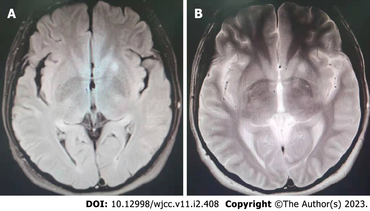
Figure 3 Brain magnetic resonance images of this patient.
A: T2-weighted fluid-attenuated inversion recovery showed slightly elevated signals within the left basal ganglia area; B: T2-Weighted scans showed slightly elevated signals within the left basal ganglia area.
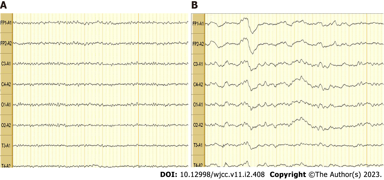
Figure 4 Video electroencephalography of this patient.
A: Wakefulness and eyes closed; B: Sleep state: Mildly abnormal electroencephalography. Multiple slow wave bursts during wakefulness. There may have been interference artifacts.
- Citation: Kong DL. Anti-leucine-rich glioma inactivated protein 1 encephalitis with sleep disturbance as the first symptom: A case report and review of literature. World J Clin Cases 2023; 11(2): 408-416
- URL: https://www.wjgnet.com/2307-8960/full/v11/i2/408.htm
- DOI: https://dx.doi.org/10.12998/wjcc.v11.i2.408


