Copyright
©The Author(s) 2023.
World J Clin Cases. Jan 16, 2023; 11(2): 401-407
Published online Jan 16, 2023. doi: 10.12998/wjcc.v11.i2.401
Published online Jan 16, 2023. doi: 10.12998/wjcc.v11.i2.401
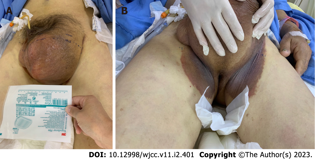
Figure 1 Left-side giant inguinoscrotal hernia.
A: Huge irreducible inguinoscrotal hernia with the penis buried within the enlarged scrotum; B: Ecchymosis formation in the bilateral inguinal region.
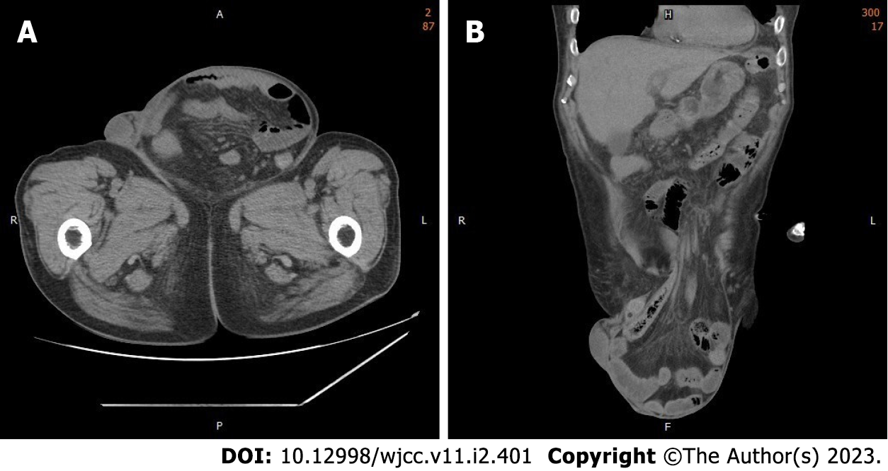
Figure 2 Abdominal computed tomography scan.
A: Computed tomography (CT) of the abdomen (axial section); B: CT scan of the abdomen (coronal section) revealing a large left-side inguinal hernia containing small bowel loops as well as the colon.
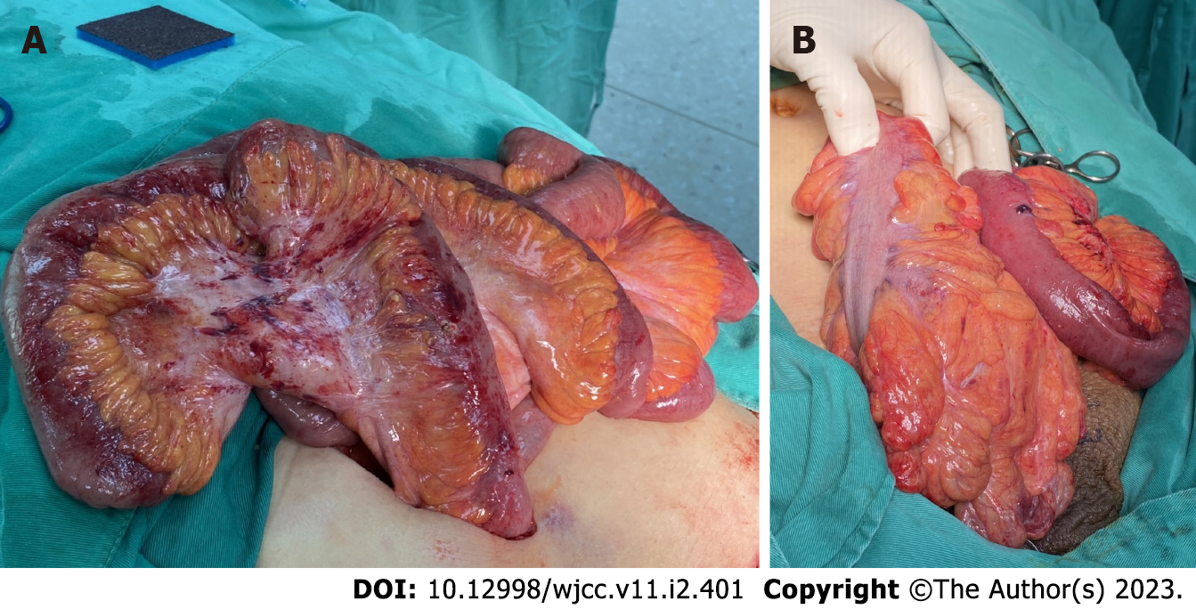
Figure 3 Intraoperative findings.
A: Hernial contents are grossly inflamed, with mild swelling and an erythematous appearance; B: The ileum and sigmoid colon from the hernial sac with relatively good perfusion.
- Citation: Liu SH, Yen CH, Tseng HP, Hu JM, Chang CH, Pu TW. Repair of a giant inguinoscrotal hernia with herniation of the ileum and sigmoid colon: A case report. World J Clin Cases 2023; 11(2): 401-407
- URL: https://www.wjgnet.com/2307-8960/full/v11/i2/401.htm
- DOI: https://dx.doi.org/10.12998/wjcc.v11.i2.401


