Copyright
©The Author(s) 2023.
World J Clin Cases. Jan 16, 2023; 11(2): 394-400
Published online Jan 16, 2023. doi: 10.12998/wjcc.v11.i2.394
Published online Jan 16, 2023. doi: 10.12998/wjcc.v11.i2.394
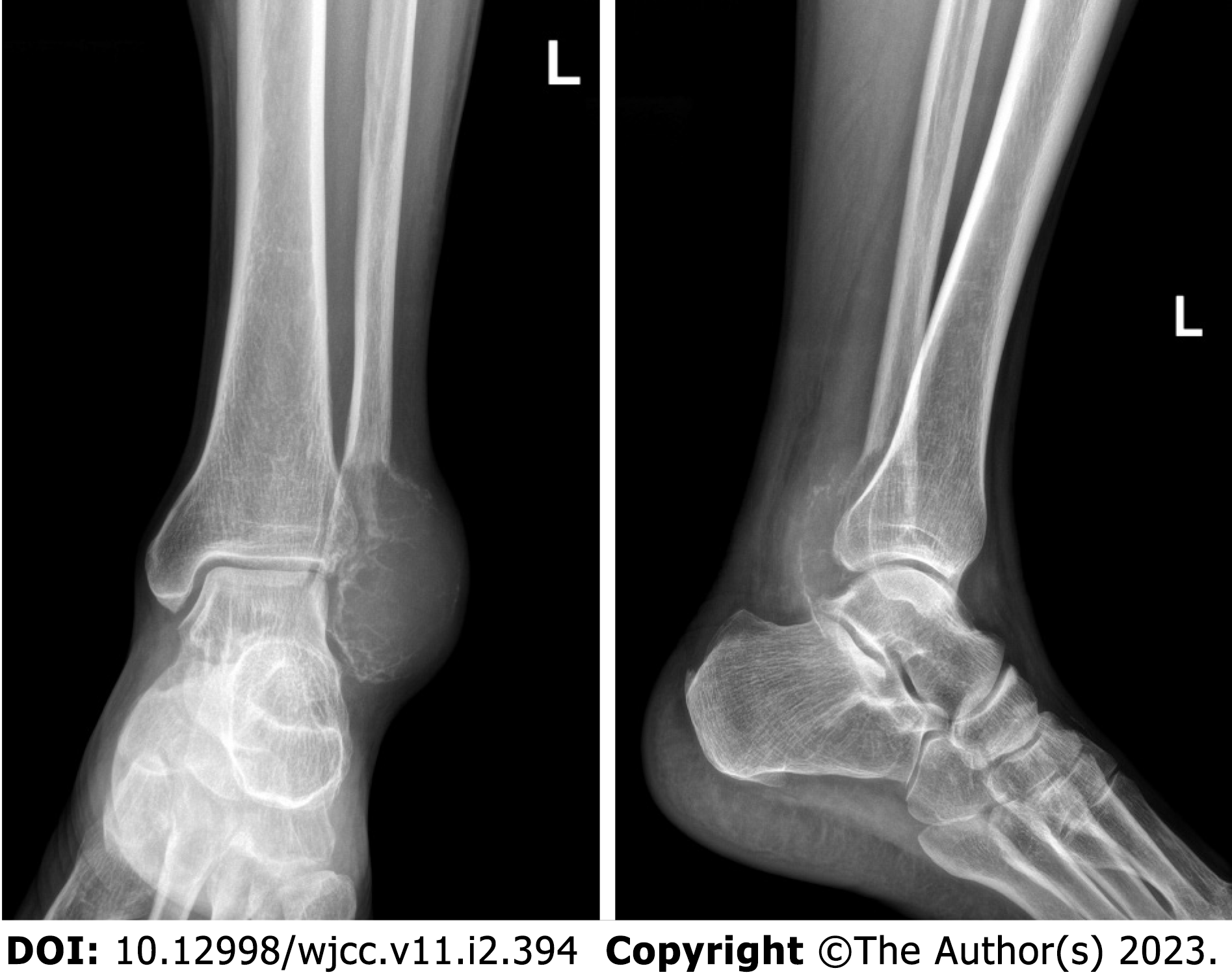
Figure 1 Radiographs showed expansile lesion with soap bubble appearance.
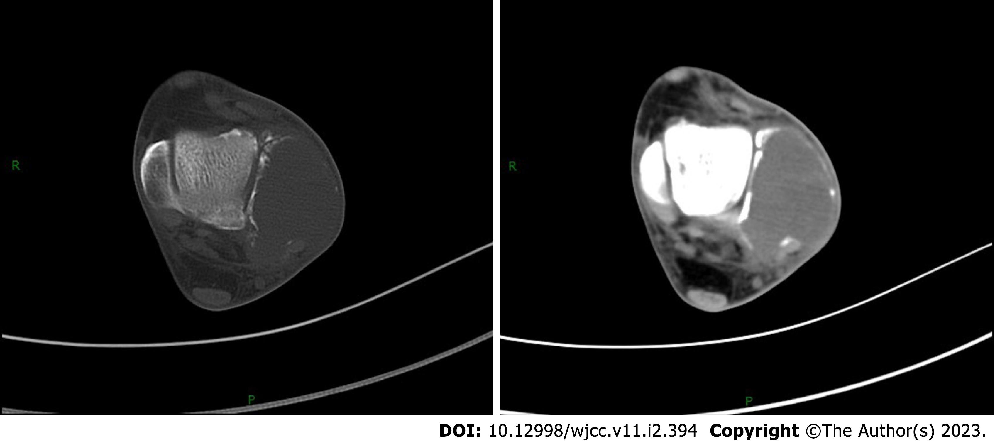
Figure 2 Computed tomography showed the distal lateral cortex of the fibula was invaded.
Figure 3 Magnetic resonance imaging scan showed soft tissue around the lateral malleolus was also invaded.
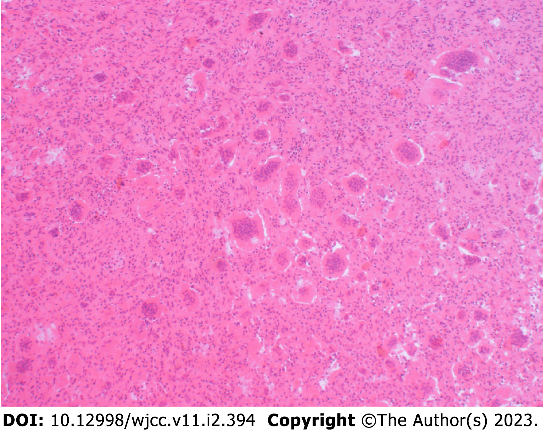
Figure 4 Pathologically, the tumor comprised mononuclear stromal cells and multinuclear giant cells.
Original magnification: × 100.
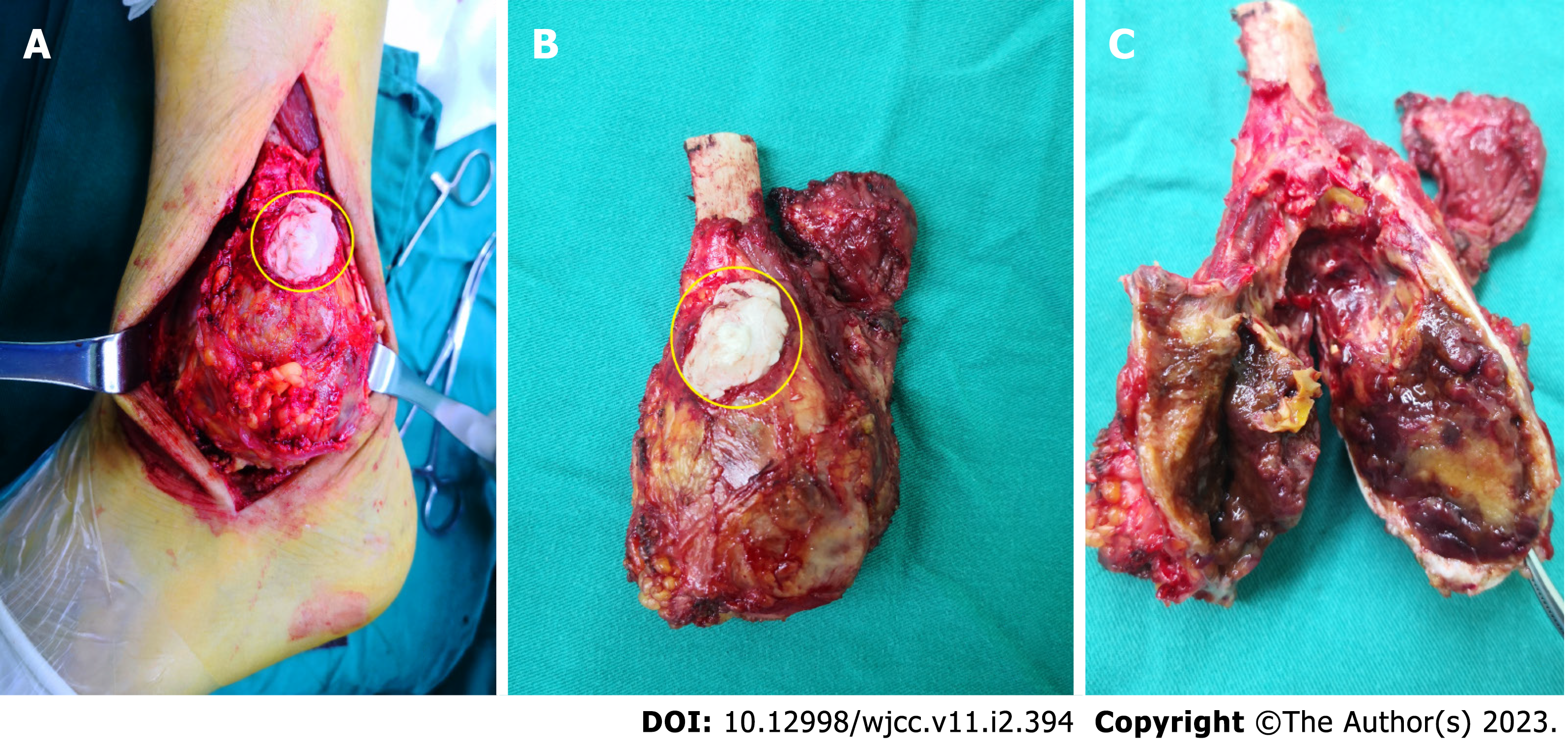
Figure 5 The macroscopic piece for the tumor: fragile, yellowish-brown tumor tissue.
A: Tumor in vivo; B: Tumor in vitro; C: Macroscopic bisection of tumor (yellow circles indicates the bone cement).
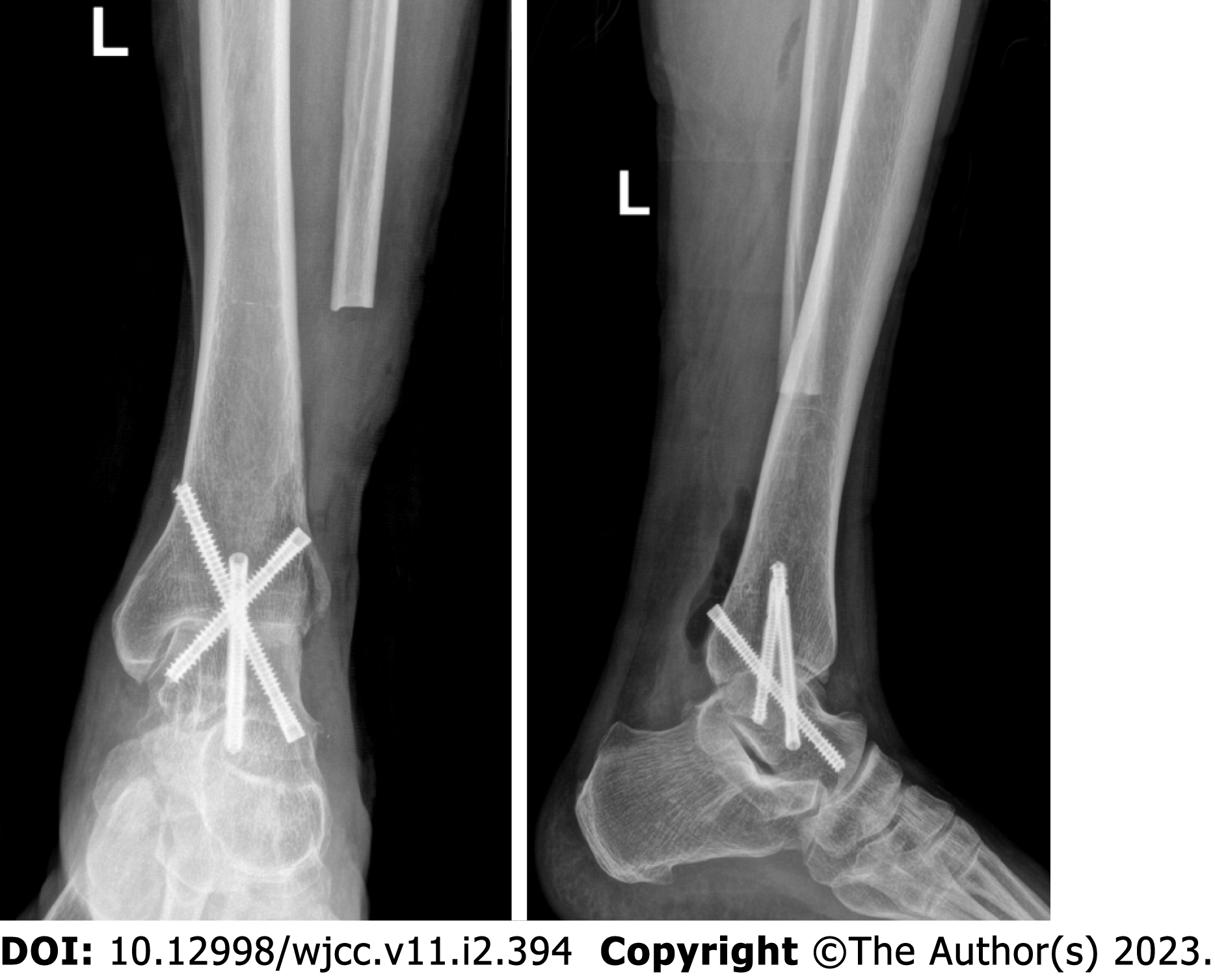
Figure 6 Postoperative X ray showed the tibial talar joint was secured with three screws.
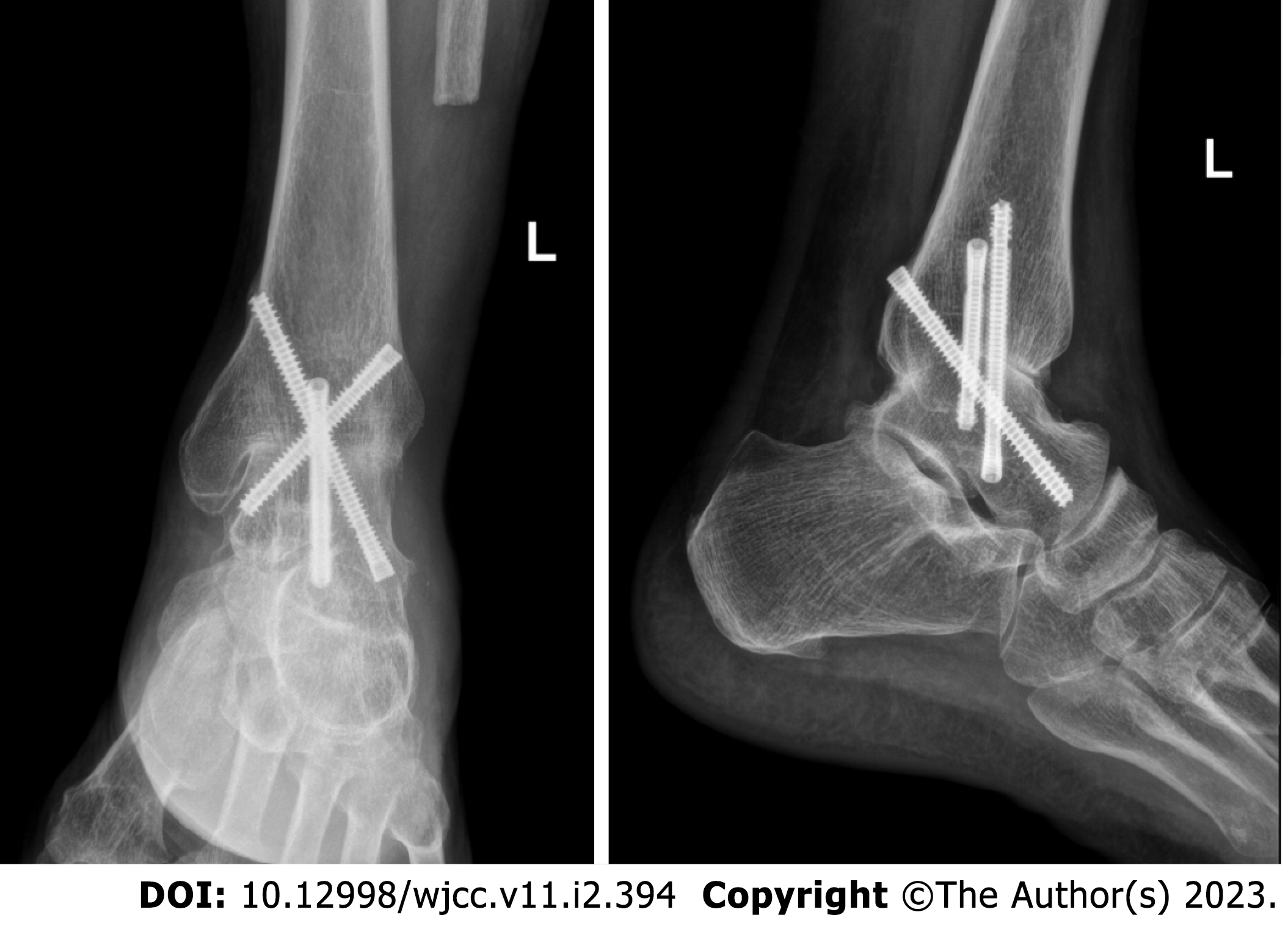
Figure 7 X-ray 2 years after the operation showed osseous fusion of the tibial talus joint.
- Citation: Fan QH, Long S, Wu XK, Fang Q. Management of a rare giant cell tumor of the distal fibula: A case report. World J Clin Cases 2023; 11(2): 394-400
- URL: https://www.wjgnet.com/2307-8960/full/v11/i2/394.htm
- DOI: https://dx.doi.org/10.12998/wjcc.v11.i2.394


