Copyright
©The Author(s) 2023.
World J Clin Cases. Jan 16, 2023; 11(2): 342-356
Published online Jan 16, 2023. doi: 10.12998/wjcc.v11.i2.342
Published online Jan 16, 2023. doi: 10.12998/wjcc.v11.i2.342
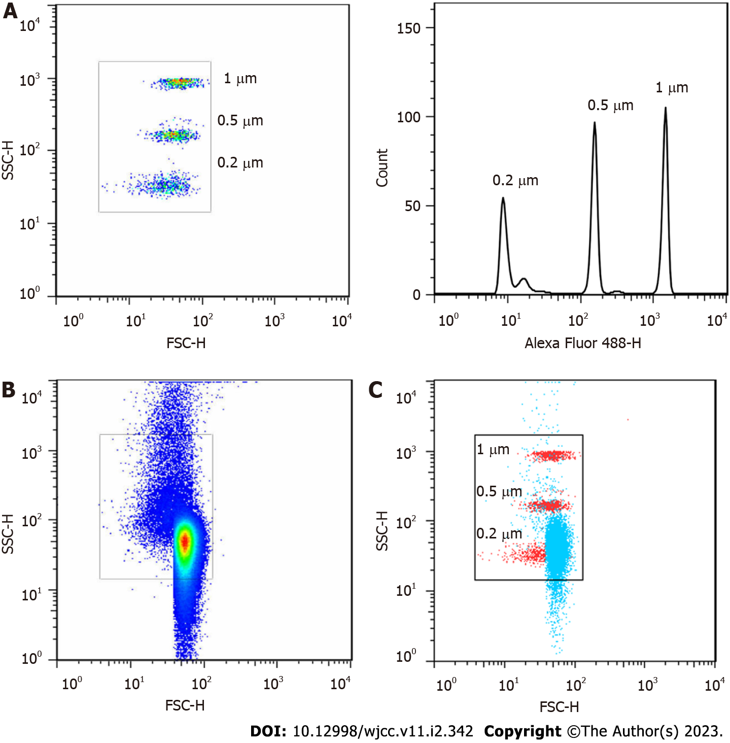
Figure 1 Gating strategy for microvesicles and characterization of microvesicles isolated from human plasma.
A: Gating strategy for microvesicles (MVs) based on calibration particles; B: Representative dot plots demonstrating the concentration of MVs isolated from human plasm; C: The merged dot plots of MVs isolated from human plasm (blue) and calibration particles (red).
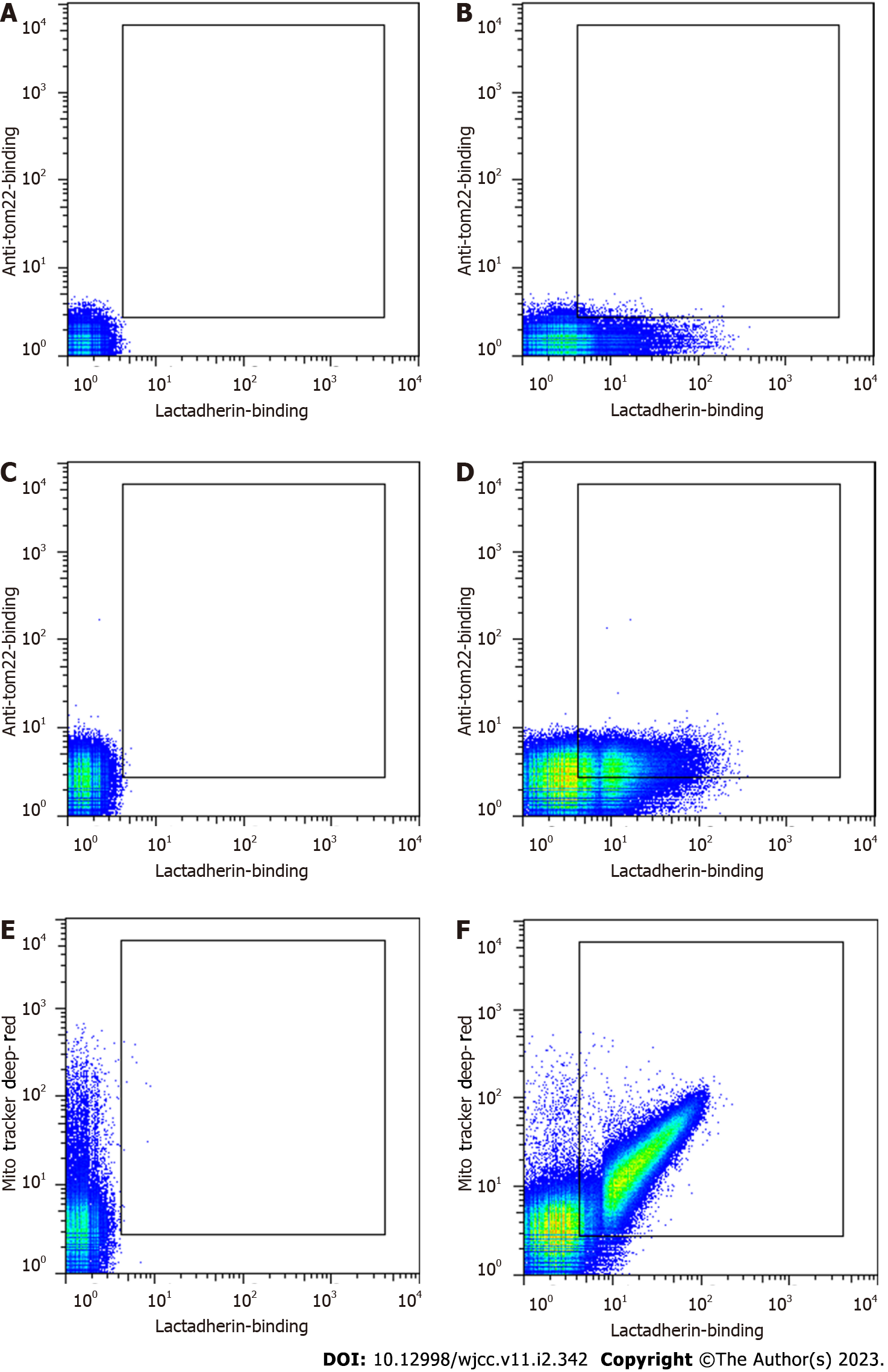
Figure 2 Representative dot plots of circulating microvesicles from a patient with sepsis.
Microvesicles (MVs) were labelled and analysed by flow cytometry. A: Unlabelled MVs (autofluorescence); B: MVs labelled with lactadherin-FITC; C: MVs labelled with anti-tom22-APC; D: MVs double stained with lactadherin-FITC and anti-tom22-APC; E: MVs labelled with MitoTracker Deep Red; F: MVs double stained with lactadherin-FITC and MitoTracker Deep Red.
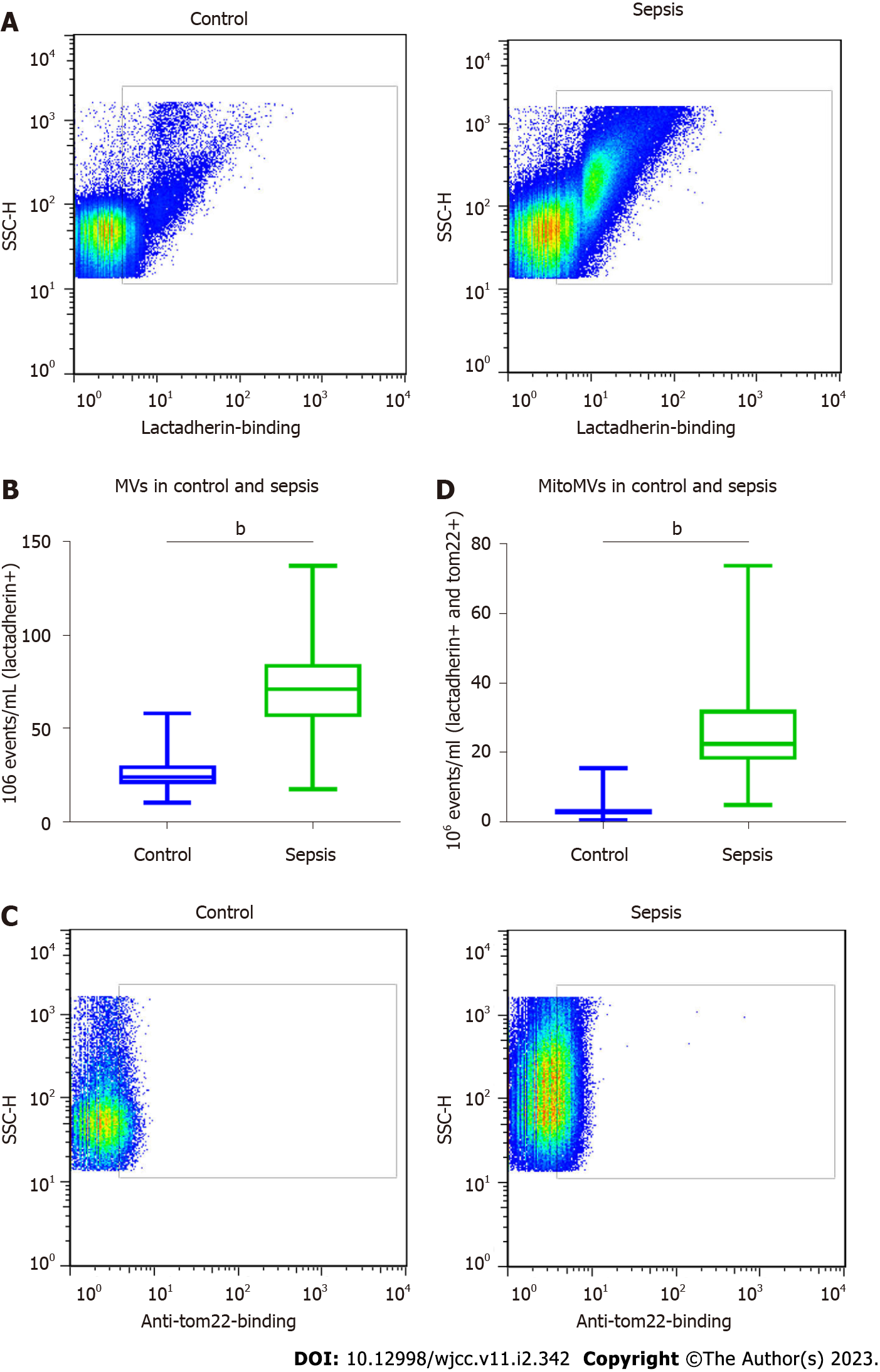
Figure 3 Flow cytometric analysis of microvesicles and microvesicles carrying mitochondrial content.
A: Representative dot plots of microvesicles (MVs) in patients with sepsis and healthy controls; B: Numbers of MVs in 19 patients with sepsis and 20 healthy controls; C: Representative dot plots of the microvesicles carrying mitochondrial content (mitoMVs) within the lactadherin+ population in patients with sepsis and healthy controls; D: Numbers of mitoMVs within the lactadherin+ population in 19 patients with sepsis and 20 healthy controls.
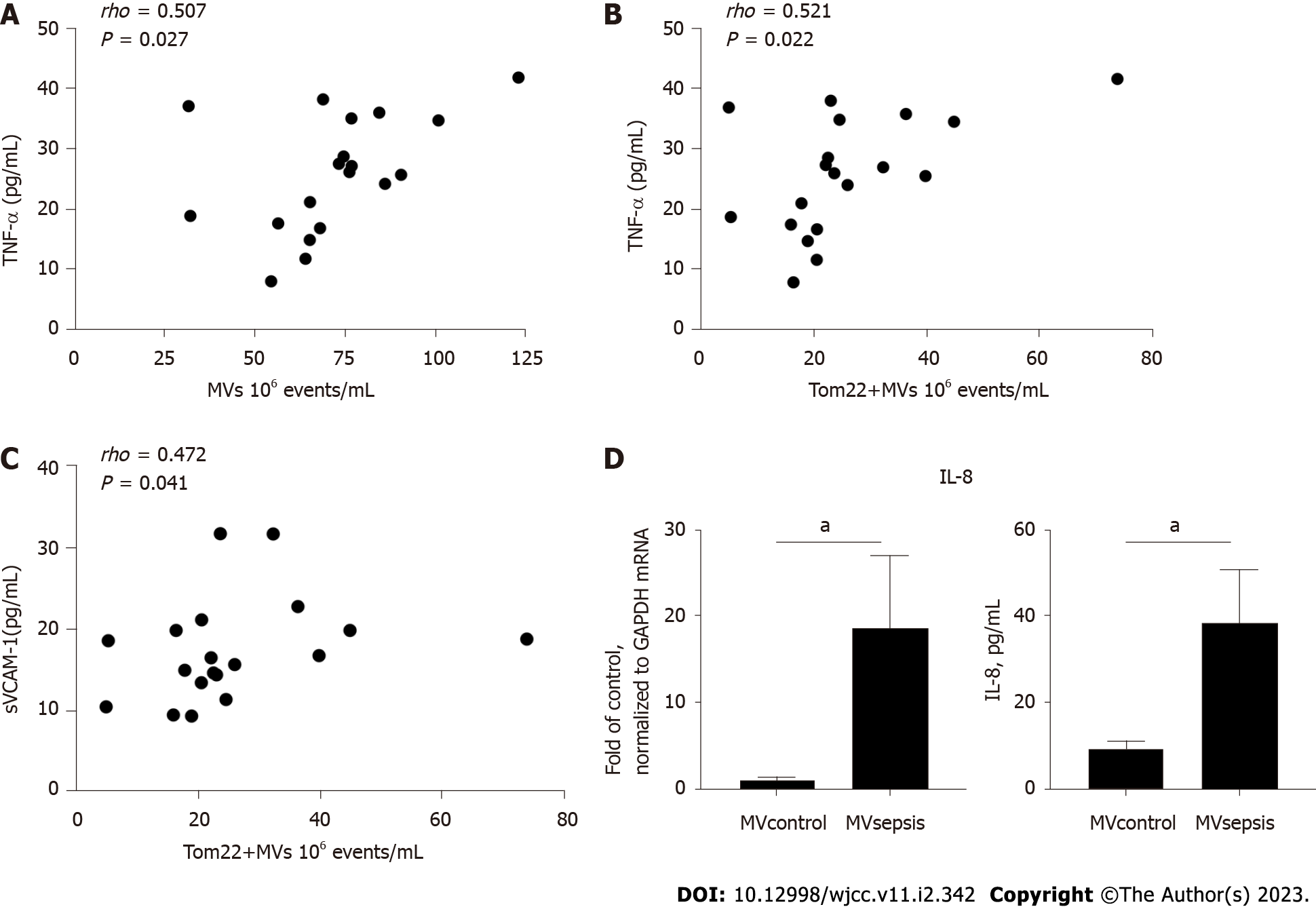
Figure 4 Association between tumour necrosis factor-α, soluble vascular cell adhesion molecule-1 and microvesicles that were isolated from the plasma of patients with sepsis and induction of interleukin-8 in human umbilical vein endothelial cells.
TNF-α: tumour necrosis factor-α; sVCAM-1: soluble vascular cell adhesion molecule-1; MVsepsis: microvesicles that were isolated from the plasma of septic patients; IL-8: interleukin-8.
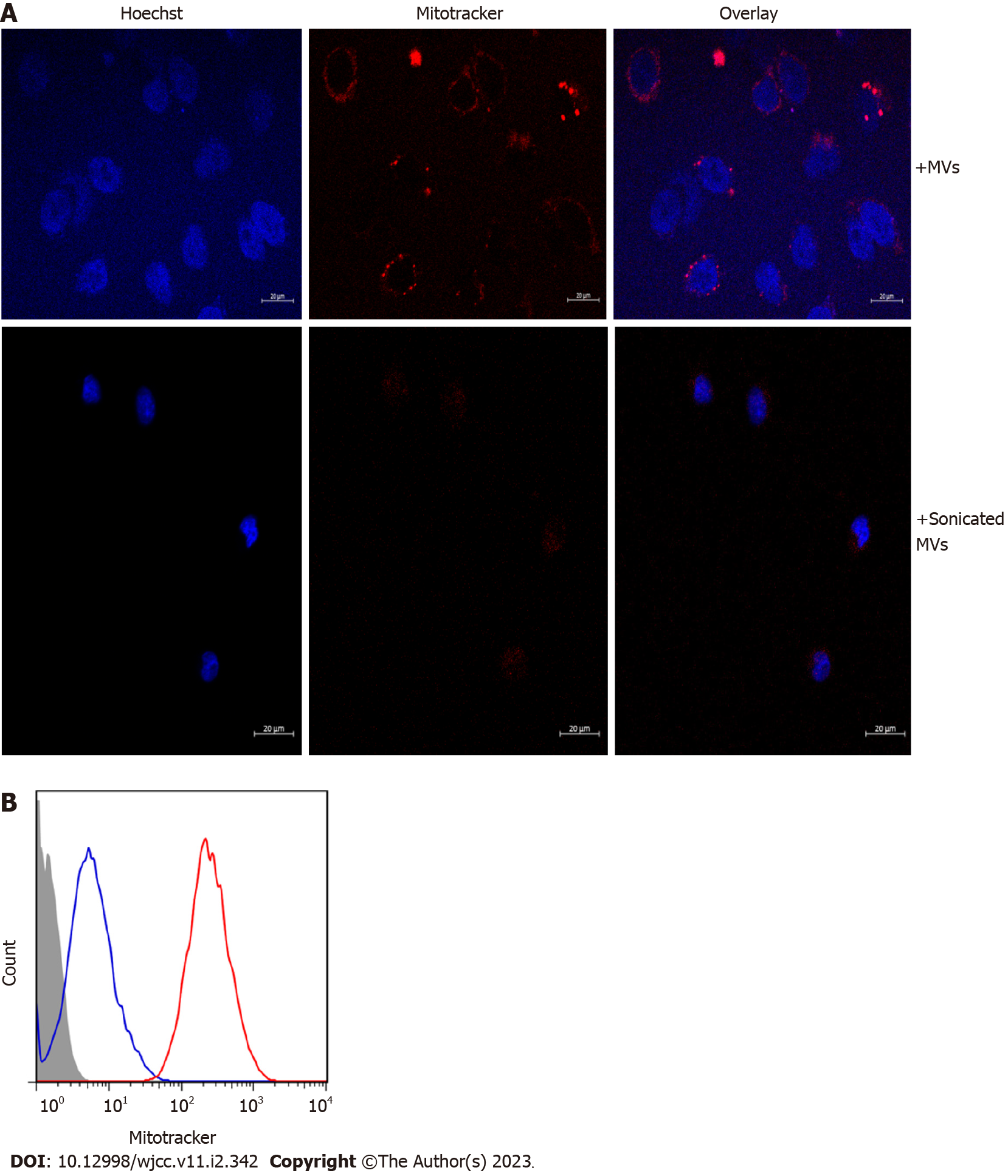
Figure 5 Uptake of microvesicles carrying mitochondrial content by human umbilical vein endothelial cells before and after ultrasonic treatment.
Microvesicles (MVs) were isolated from the plasma of patients with sepsis and labelled with MitoTracker Deep Red. A: Human umbilical vein endothelial cells (HUVECs) were incubated with the labelled MVs and treated or not treated with sonication. Nuclei were stained by Hoechst; B: Flow cytometric analysis of HUVECs after incubation with the labelled MVs either treated (blue) or not (red) treated with sonication.
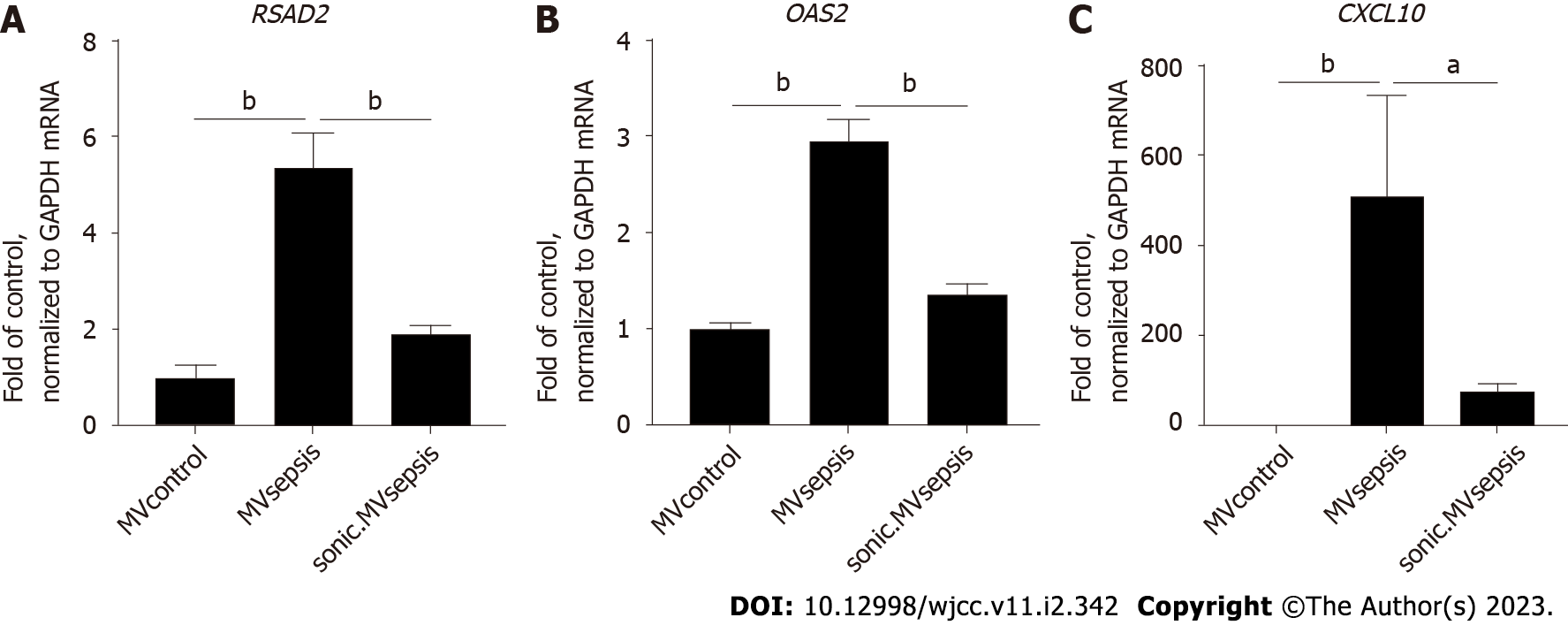
Figure 6 mRNA expression in human umbilical vein endothelial cells after stimulation with circulating microvesicles.
MVcontrol: microvesicles that were isolated from the plasma of healthy controls; MVsepsis: microvesicles that were isolated from the plasma of septic patients; sonic. MVsepsis: MVsepsis that were treated by sonication.
- Citation: Zhang HJ, Li JY, Wang C, Zhong GQ. Microvesicles with mitochondrial content are increased in patients with sepsis and associated with inflammatory responses. World J Clin Cases 2023; 11(2): 342-356
- URL: https://www.wjgnet.com/2307-8960/full/v11/i2/342.htm
- DOI: https://dx.doi.org/10.12998/wjcc.v11.i2.342


