Copyright
©The Author(s) 2023.
World J Clin Cases. Jul 6, 2023; 11(19): 4698-4706
Published online Jul 6, 2023. doi: 10.12998/wjcc.v11.i19.4698
Published online Jul 6, 2023. doi: 10.12998/wjcc.v11.i19.4698
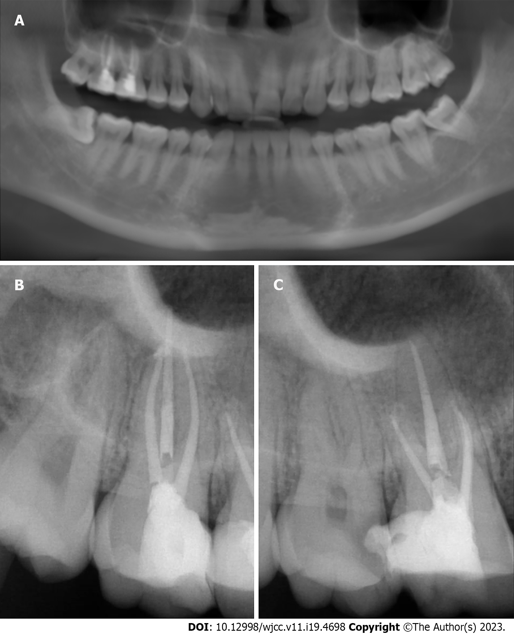
Figure 1 Panoramic and periapical films of teeth #16 and #17.
A: Panoramic of teeth #16 and #17; B and C: Periapical films of teeth #16 and #17, both #16 and #17 had undergone successful root canal treatment.
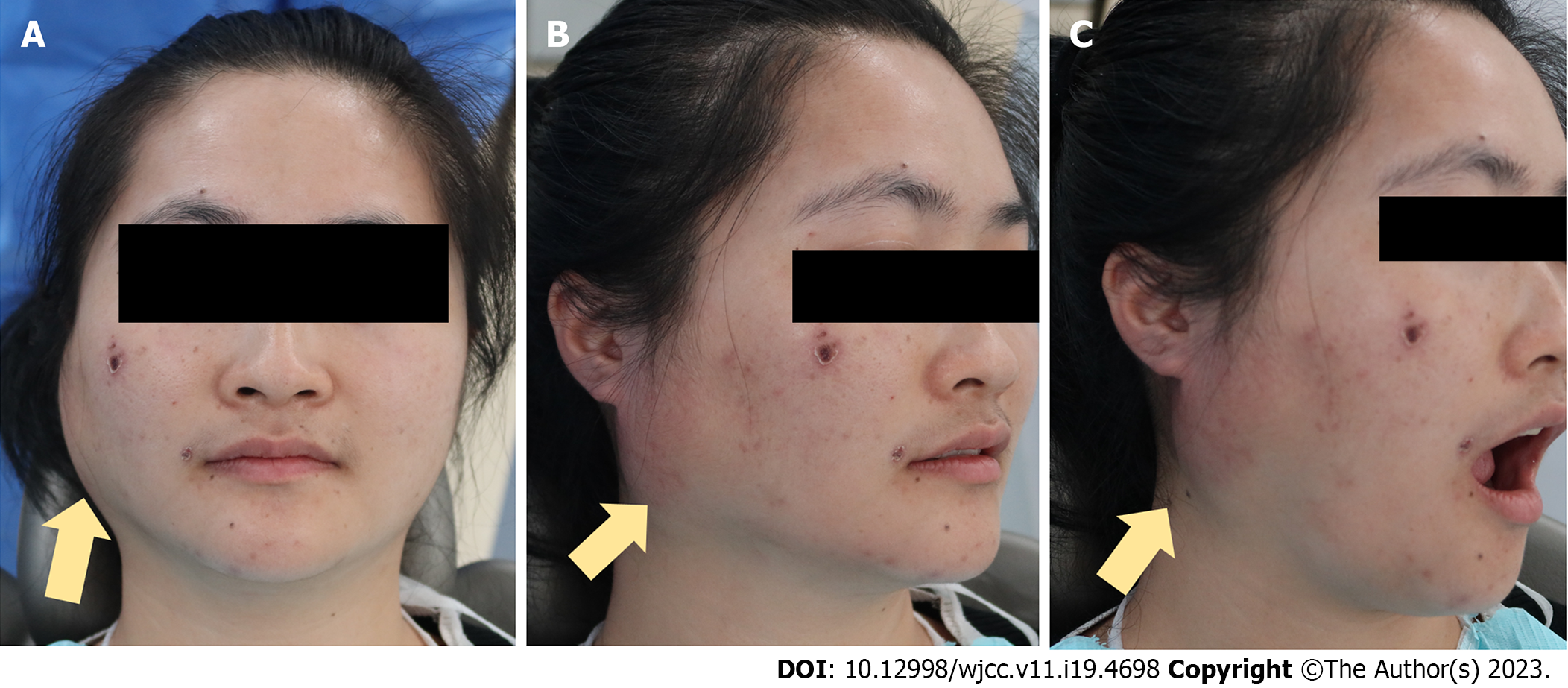
Figure 2 Subcutaneous emphysema is clearly evident in the right retromandibular angle.
The skin temperature rose and the skin became red. Mouth-opening was not restricted and the patient felt no pain in the region of swelling. Yellow arrows: Subcutaneous emphysema. A: Front view; B: Profile view; C: Profile view while mouth opening.
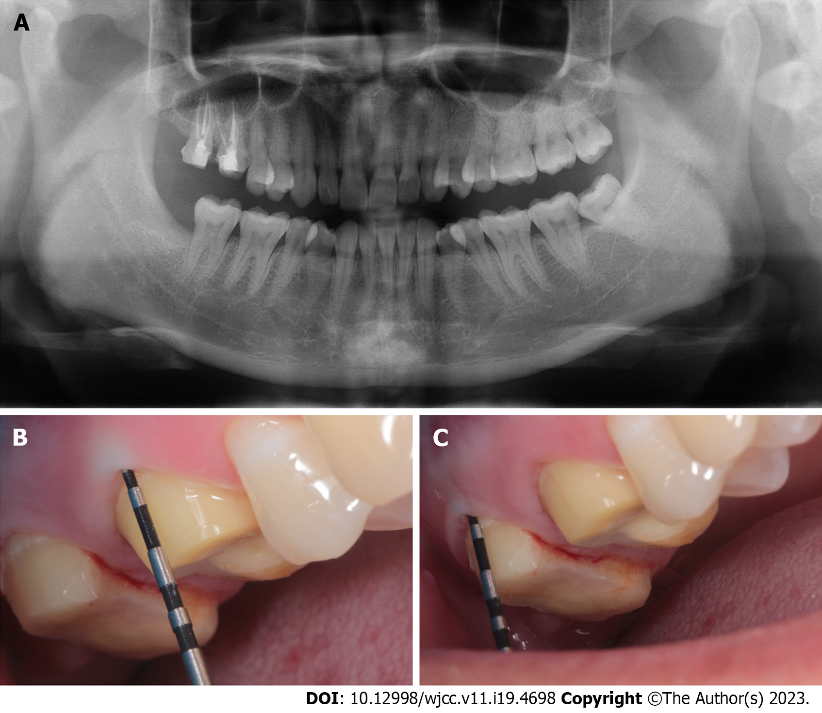
Figure 3 Panoramic imaging and gingival sulcus depth probing were performed after emphysema developed.
A: Panoramic imaging was performed after emphysema developed; B and C: Gingival sulcus depth probing were performed after emphysema developed. Gingival sulcus depth pocket probing was normal (buccal lateral: #16: 2 mm; #17: 2.8 mm). No significant anomaly was found.
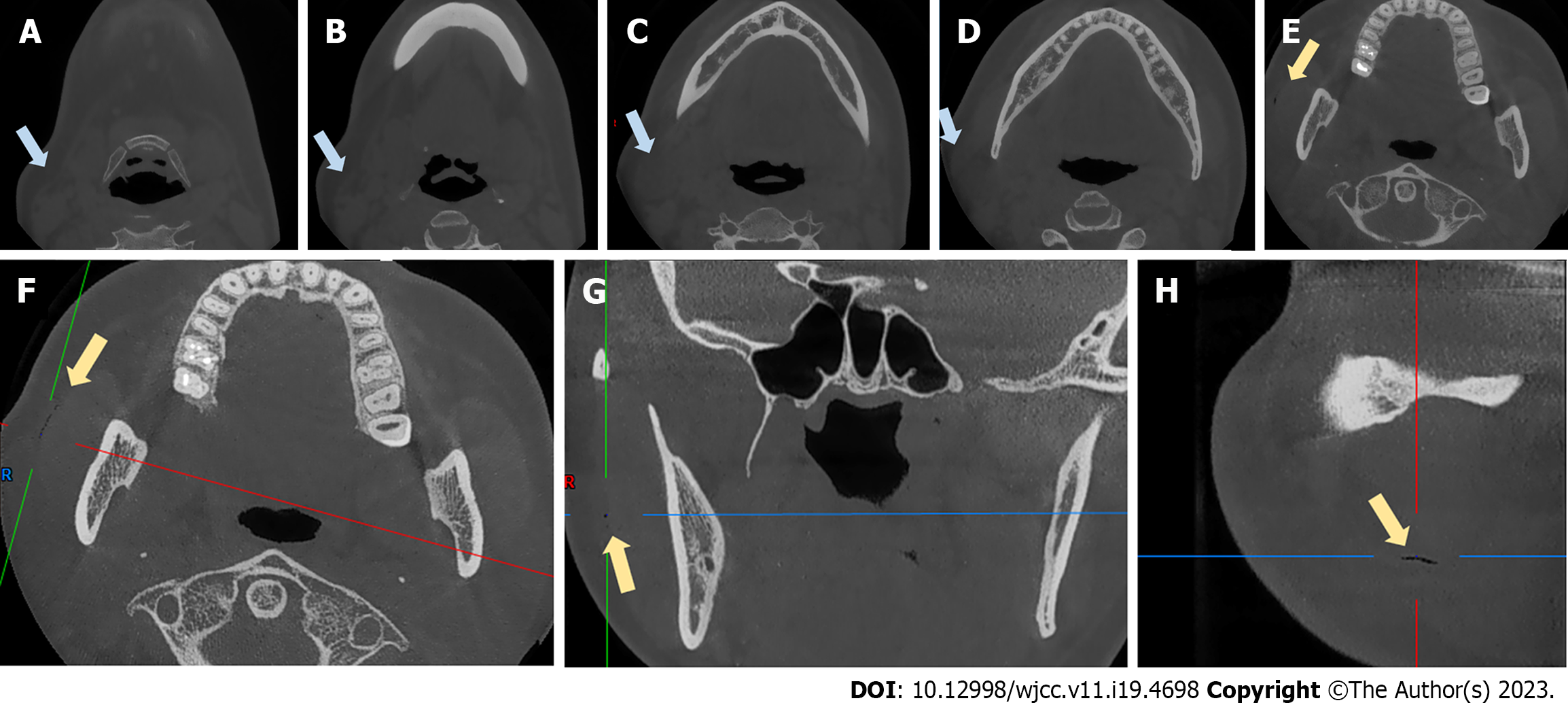
Figure 4 Cone-beam computed tomography scans obtained after emphysema developed.
A-E: Coronal views of the swelling; F-H: An air-filled fissure between the masseter muscle and soft tissue. Blue arrows: Swelling. Yellow arrows: The fissure.
Figure 5 After 3 d of intravenous drip, the swelling of the right retromandibular angle disappeared.
A: Front view; B: Profile view.
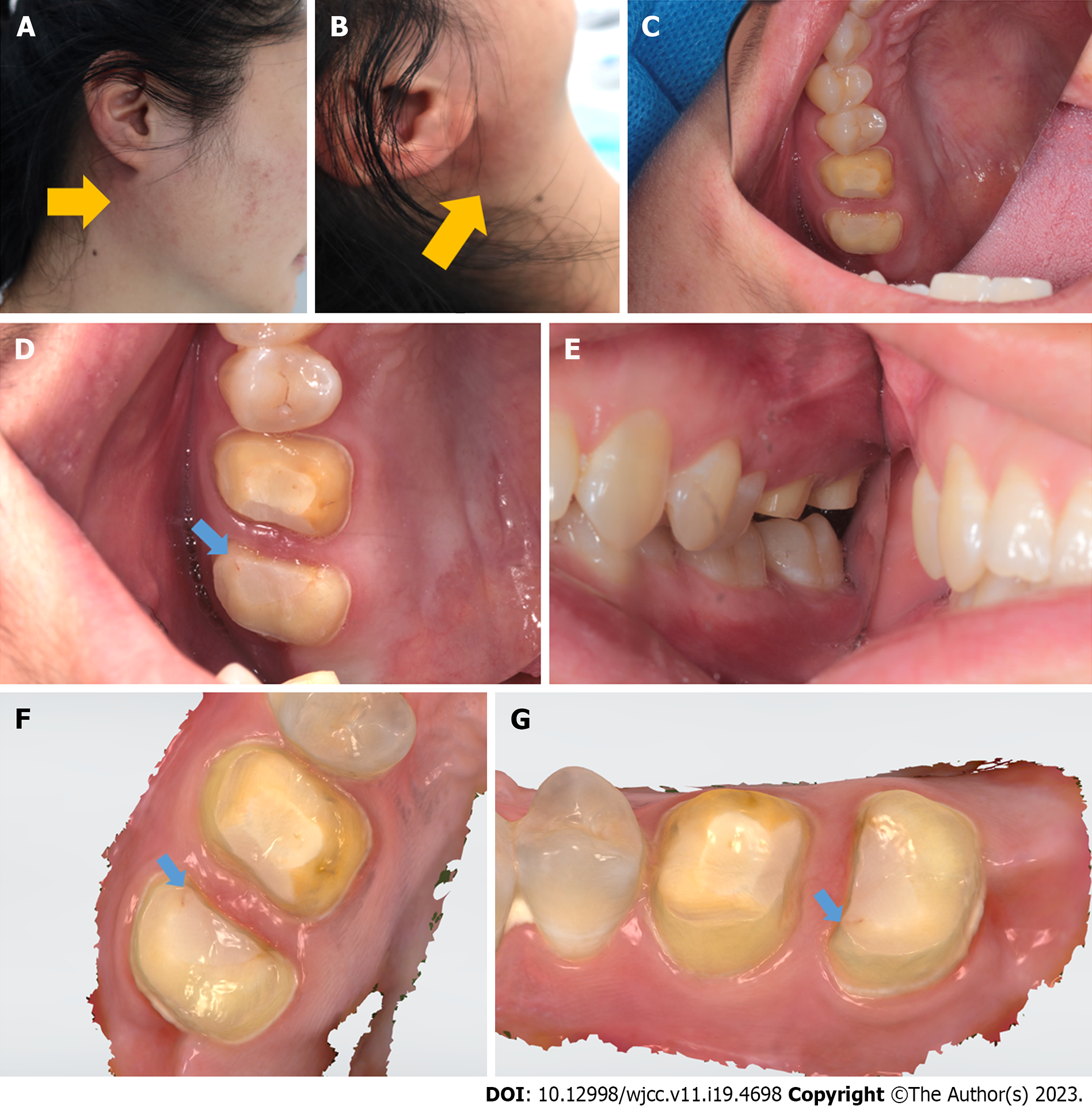
Figure 6 The symptoms were similar to those on the first occasion.
A and B: The emphysema was in the retromandibular area; the yellow arrows indicate the swelling; C-E: The teeth and gingiva were normal and healthy; F and G: Optical scans acquired using an oral scanner (3Shape, Copenhagen, Denmark) also revealed very good general health. However, beginning on the occlusion surface of #17, a subfissure line was found between the tooth and resin (blue arrow). Grinding at this point triggered subcutaneous emphysema in the retromandibular area.
- Citation: Bai YP, Sha JJ, Chai CC, Sun HP. With two episodes of right retromandibular angle subcutaneous emphysema during right upper molar crown preparation: A case report. World J Clin Cases 2023; 11(19): 4698-4706
- URL: https://www.wjgnet.com/2307-8960/full/v11/i19/4698.htm
- DOI: https://dx.doi.org/10.12998/wjcc.v11.i19.4698


