Copyright
©The Author(s) 2023.
World J Clin Cases. Jul 6, 2023; 11(19): 4692-4697
Published online Jul 6, 2023. doi: 10.12998/wjcc.v11.i19.4692
Published online Jul 6, 2023. doi: 10.12998/wjcc.v11.i19.4692
Figure 1 Clinical manifestations of the patient before and after treatment.
A: Initial clinical presentation. Moderate ptosis (yellow lines), suspected edema inside bilateral upper eyelids and shallow glabellar wrinkles were observed; B: Seven days after treatment of minocycline and ketotifen. Erythema (yellow arrow) triggered by biopsy and obvious improvement of ptosis and deepened glabellar wrinkles; C: 40 days after treatment of minocycline and ketotifen. The bilateral eyelids basically recovered to the pre-illness state.
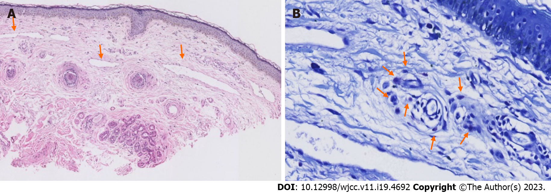
Figure 2 Biopsy specimen from patient's upper eyelid.
A: Hematoxylin–eosin staining (×40) showing edema and dilated lymphatics in dermis (orange arrows). B: Increased infiltration of mast cell (orange arrows) in toluidine blue (×400).
- Citation: Na J, Wu Y. Morbihan disease misdiagnosed as senile blepharoptosis and successfully treated with short-term minocycline and ketotifen: A case report. World J Clin Cases 2023; 11(19): 4692-4697
- URL: https://www.wjgnet.com/2307-8960/full/v11/i19/4692.htm
- DOI: https://dx.doi.org/10.12998/wjcc.v11.i19.4692


