Copyright
©The Author(s) 2023.
World J Clin Cases. Jul 6, 2023; 11(19): 4684-4691
Published online Jul 6, 2023. doi: 10.12998/wjcc.v11.i19.4684
Published online Jul 6, 2023. doi: 10.12998/wjcc.v11.i19.4684
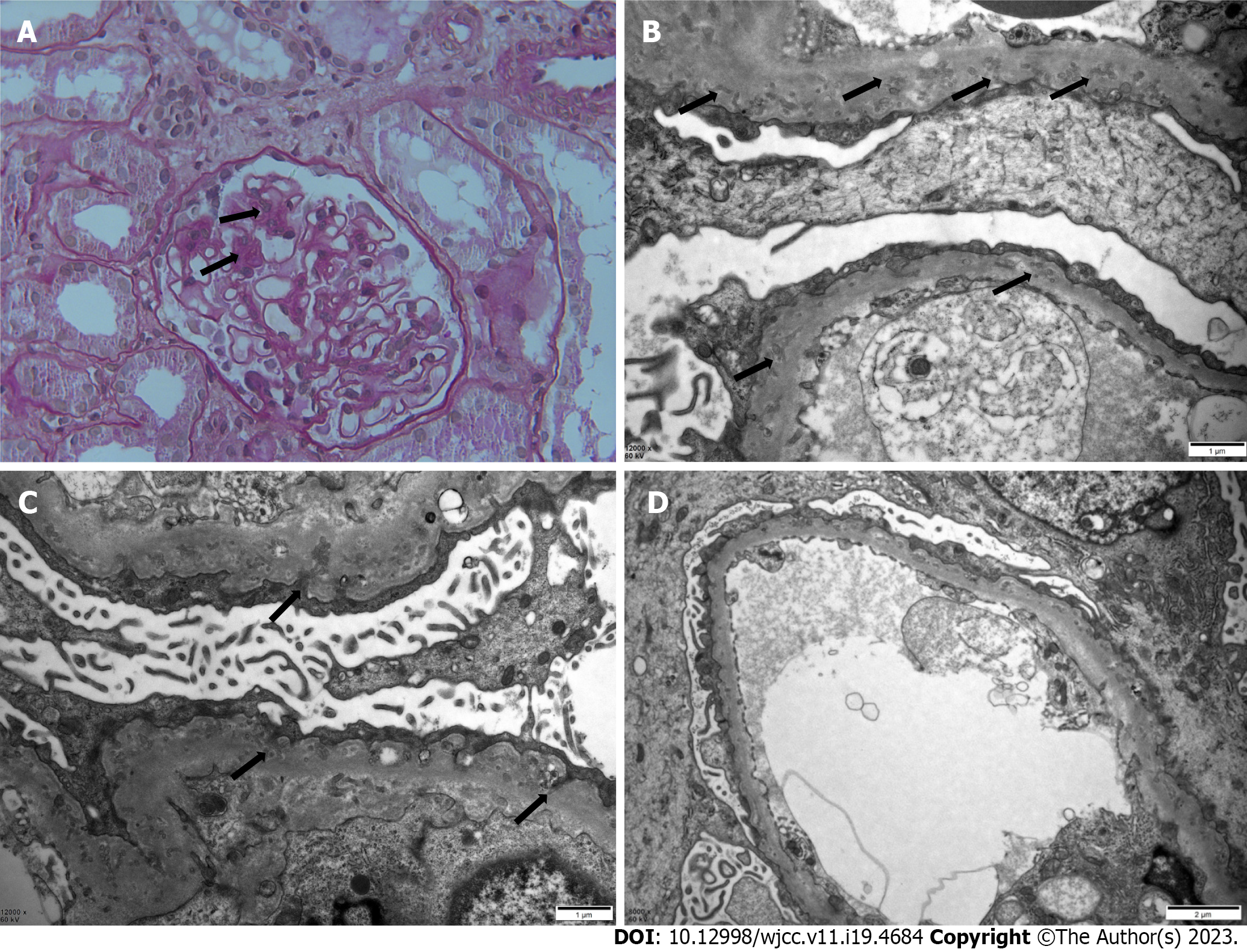
Figure 1 Kidney biopsy findings.
A: Light microscopic findings on periodic acid-Schiff staining. The mesangial area showed mild focal segmental proliferation and matrix thickening (arrows). The basement membrane is mildly thickened (original magnification, × 400); B: Electron microscopy of kidney biopsy specimen. Thickened glomerular basement membrane (GBM) with numerous diffusely scattered microspherical or microtubular structures (arrows), characteristic of podocyte infolding glomerulopathy (original magnification, × 12000); C: Extensive podocyte foot-process effacement with infolding into the GBM (arrows) (original magnification, × 12000); D: There are no electron-dense deposits (original magnification, × 8000).
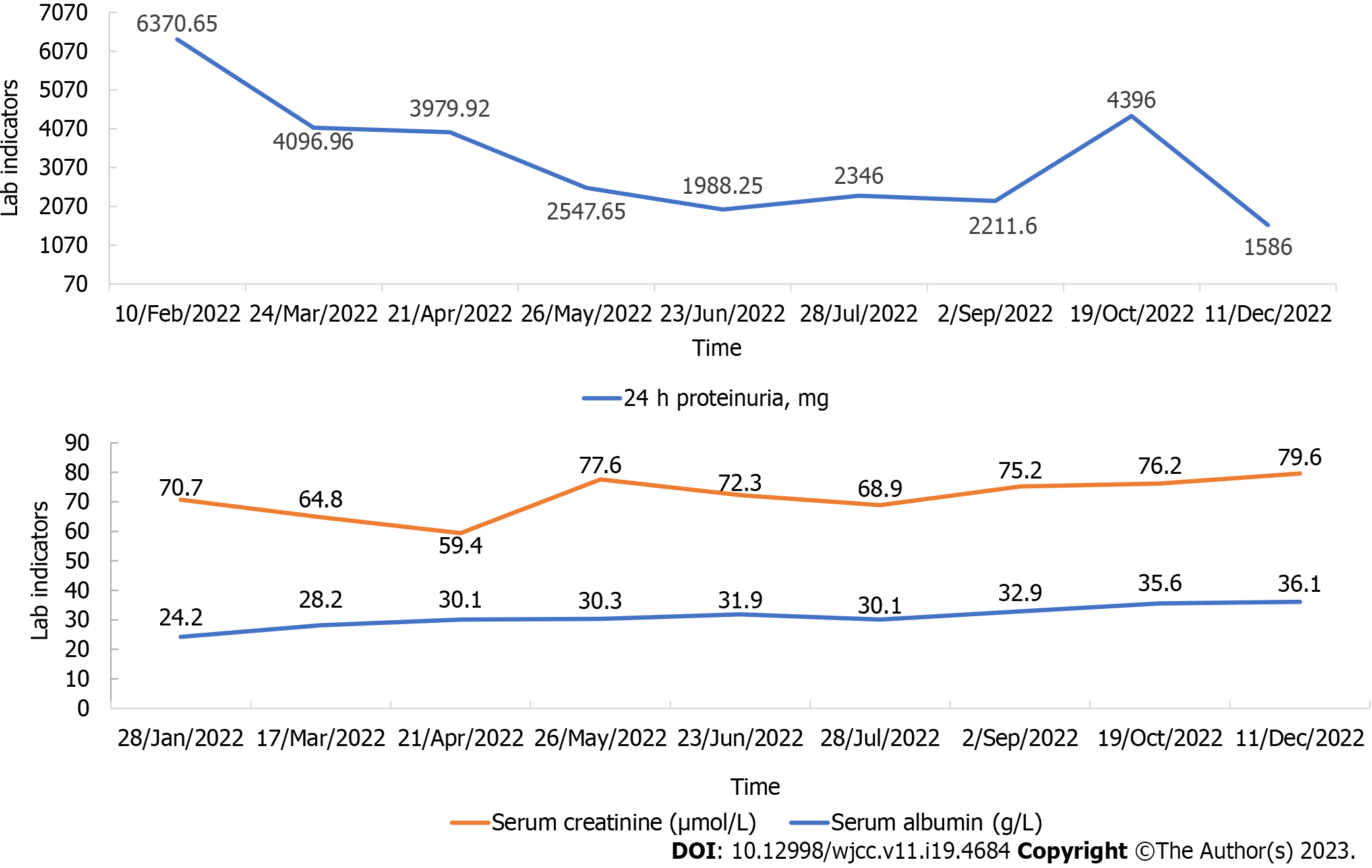
Figure 2 Changes in 24-h proteinuria (mg), serum creatinine (μmol/L), and serum albumin (g/L) over time.
- Citation: Chang MY, Zhang Y, Li MX, Xuan F. Integrated Chinese and Western medicine in the treatment of a patient with podocyte infolding glomerulopathy: A case report. World J Clin Cases 2023; 11(19): 4684-4691
- URL: https://www.wjgnet.com/2307-8960/full/v11/i19/4684.htm
- DOI: https://dx.doi.org/10.12998/wjcc.v11.i19.4684


