Copyright
©The Author(s) 2023.
World J Clin Cases. Jun 26, 2023; 11(18): 4425-4432
Published online Jun 26, 2023. doi: 10.12998/wjcc.v11.i18.4425
Published online Jun 26, 2023. doi: 10.12998/wjcc.v11.i18.4425
Figure 1 Chest X-ray of patient.
A: Chest X-ray findings on admission, increased texture in both lungs and hyperdense shadows in both lungs with an unclear border; B: Chest radiograph results at intensive care unit, bilateral lung exudation increased compared with the previous one, complicated by pneumothorax; C: Chest X-ray images at discharge. The exudate was partially absorbed in both lungs.
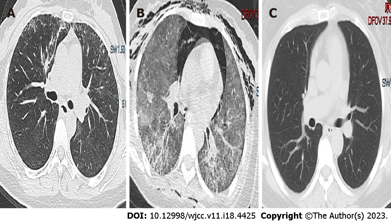
Figure 2 Chest computed tomography scans of patient.
A: Computed tomography (CT) images on admission, diffuse stripes and cords in both lungs with increased density and fuzzy edges; B: CT results in the intensive care unit, consolidation, aerothorax and ground-glass opacities in both lungs; C: CT findings at discharge, the marked resolution of ground-glass opacities and consolidation.
- Citation: Huang JJ, Zhang SS, Liu ML, Yang EY, Pan Y, Wu J. Next-generation sequencing technology for the diagnosis of Pneumocystis pneumonia in an immunocompetent female: A case report. World J Clin Cases 2023; 11(18): 4425-4432
- URL: https://www.wjgnet.com/2307-8960/full/v11/i18/4425.htm
- DOI: https://dx.doi.org/10.12998/wjcc.v11.i18.4425


