Copyright
©The Author(s) 2023.
World J Clin Cases. Jun 26, 2023; 11(18): 4419-4424
Published online Jun 26, 2023. doi: 10.12998/wjcc.v11.i18.4419
Published online Jun 26, 2023. doi: 10.12998/wjcc.v11.i18.4419
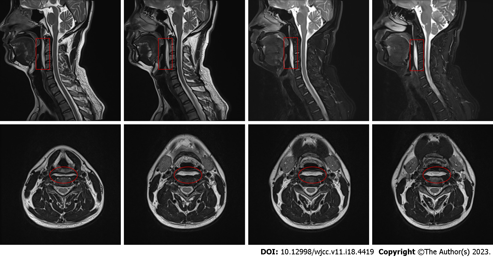
Figure 1 Sagittal section of the T2-weighted cervical magnetic resonance imaging scan.
Prevertebral effusion at the C1-C4 level (indicated with a red box); Axial (indicated with a red circle).
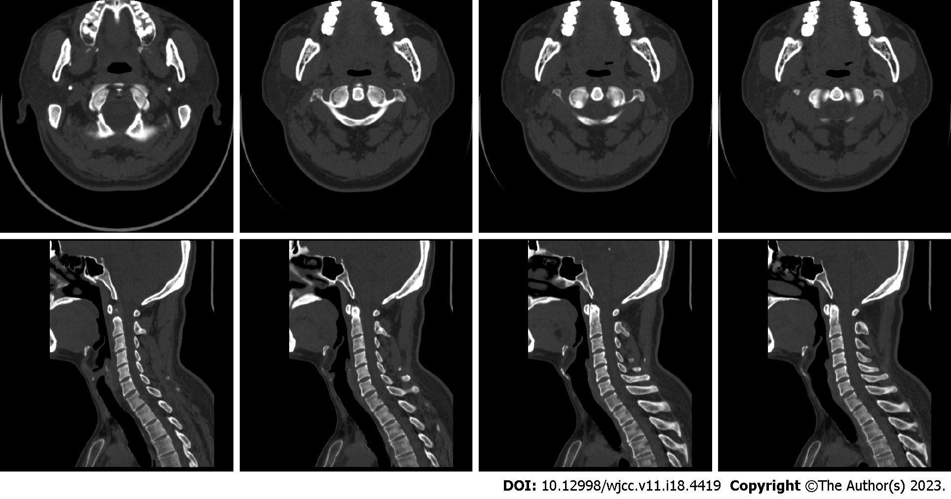
Figure 2 The computed tomography scan of the neck, which shows no calcification.
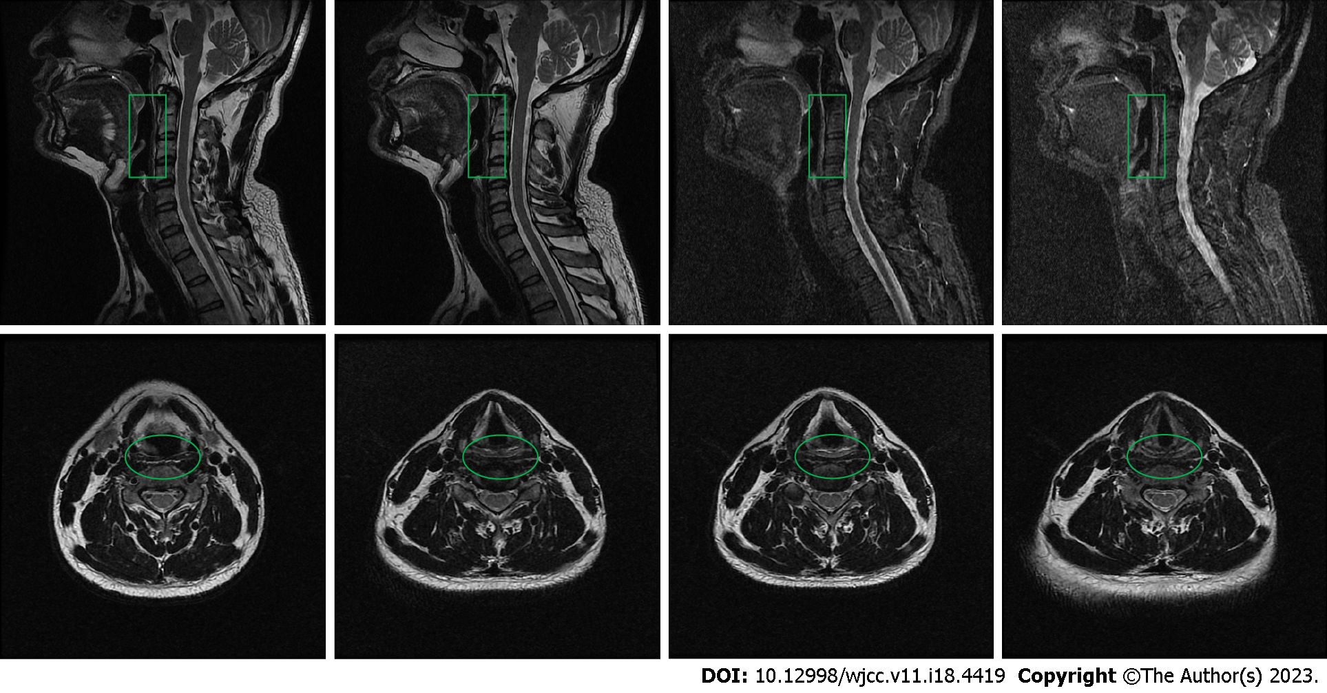
Figure 3 Sagittal and axial magnetic resonance imaging scan of the cervical spine after active treatment.
No significant prevertebral effusions at the C1-C4 level (indicated with a green box); axial (indicated with a green circle).
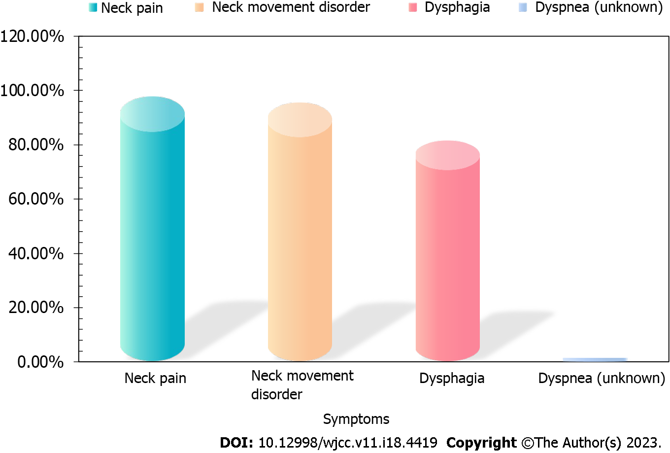
Figure 4 The graph shows the incidence rate of the symptom.
- Citation: Wu H, Liu W, Mi L, Liu Q. Acute neck tendonitis with dyspnea: A case report. World J Clin Cases 2023; 11(18): 4419-4424
- URL: https://www.wjgnet.com/2307-8960/full/v11/i18/4419.htm
- DOI: https://dx.doi.org/10.12998/wjcc.v11.i18.4419


