Copyright
©The Author(s) 2023.
World J Clin Cases. Jun 26, 2023; 11(18): 4397-4405
Published online Jun 26, 2023. doi: 10.12998/wjcc.v11.i18.4397
Published online Jun 26, 2023. doi: 10.12998/wjcc.v11.i18.4397
Figure 1 Physical examination of the sternum (manubriosternal joint) region.
A: Lateral; B: Frontal views; A and B: Reddish, slightly protruding, small, painless 3 cm × 3 cm lump in the projection of the sternum (arrows).
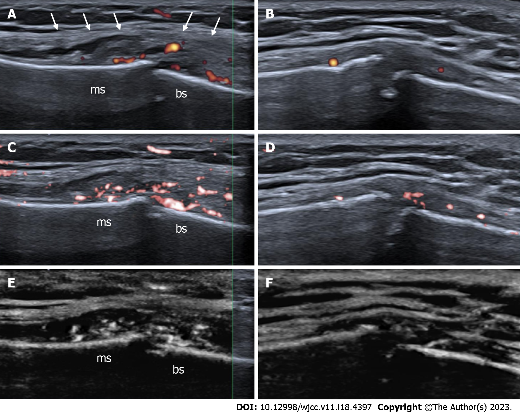
Figure 2 High-resolution ultrasound images with superb microvascular imaging of the manubriosternal joint, longitudinal views.
A: The pseudo-capsular layer is separated by intra-articular effusion and synovial hypertrophy (white arrows) with remarkable power Doppler signals; B: Four-month follow-up by power Doppler; C: Color superb microvascular imaging (SMI) visualizes slow flow vascularity more sensitive than conventional power Doppler. Active synovial inflammation with subsequent effusion formation allows suspecting of septic arthritis; D: Four-month follow-up by color SMI; E: Monochrome SMI helps to catch the eye and highlights true flow signals of small vessels in the grey map. F: Four-month follow-up by monochrome SMI. There is still mild peri- and intra-articular vascularity or arthritis and more eroded joint margins. A diagnostic ultrasound system (CANON TUS-AI800, Canon medical systems corp, Shimoishigami, Otawara-shi, Japan) equipped with a linear transducer with the following settings (14 MHz frequency) was used; ms: Manubrium of the sternum; bs: Body of the sternum.
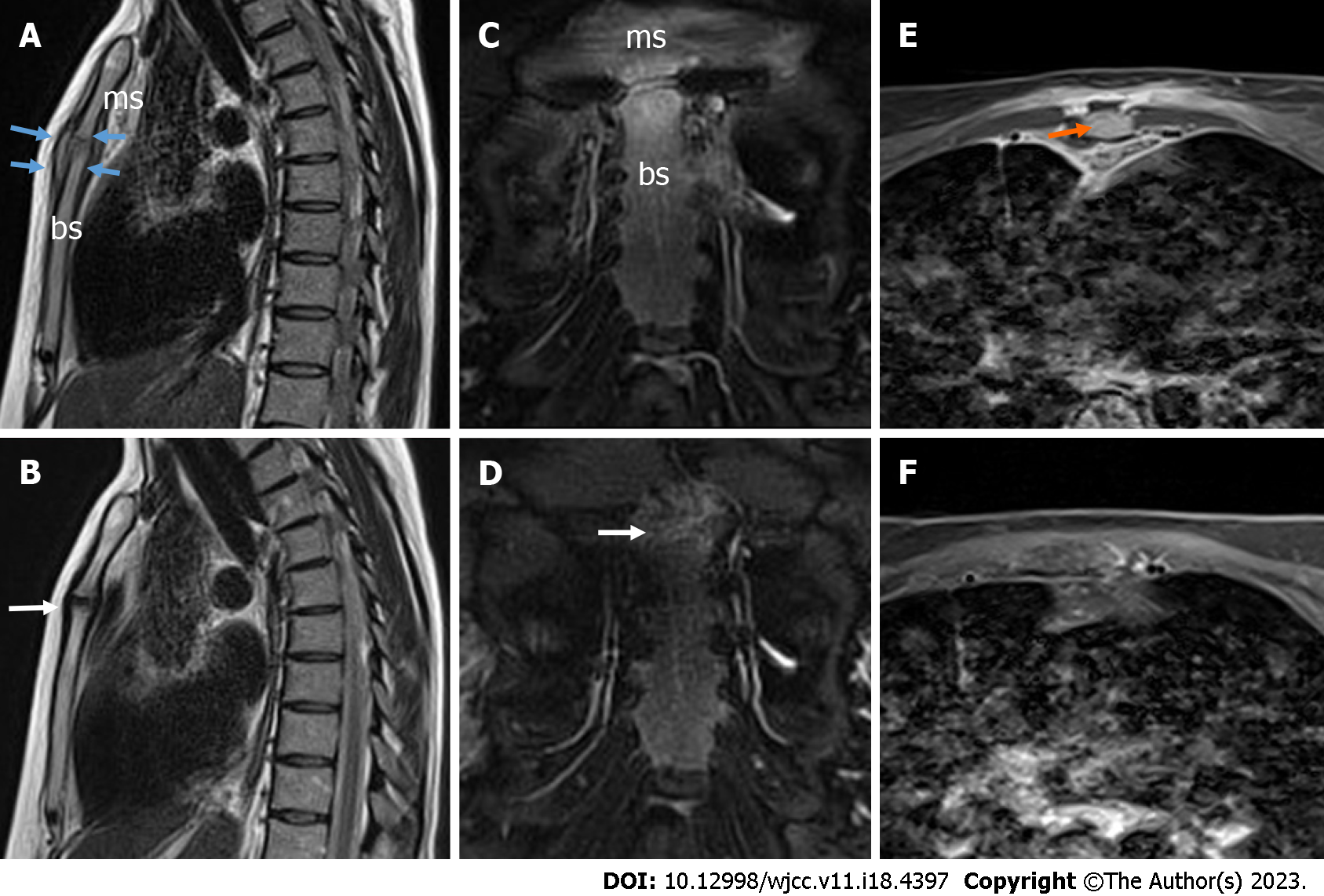
Figure 3 Magnetic resonance imaging of the septic manubriosternal joint.
A: Magnetic resonance imaging (MRI) sagittal T2-weighted blade image. The surfaces of the manubriosternal (MS) joint are eroded in the margins, the joint is enlarged, and the pseudo-capsule is thickened (blue arrows), with visible arthritis; B: MRI sagittal T2-weighted blade image. Four-month follow-up results - the surfaces of the MS joint are further eroded in the margins, perichondritis (white arrow). The pseudo-capsule is thinned; C: MRI coronal T2-weighted blade fat-suppressed image. A mild bone marrow edema is detected; D: MRI coronal T2-weighted blade fat-suppressed image. Four-month follow-up results - a very mild bone marrow edema was detected (white arrow); E: MRI axial contrast-enhanced T1-weighted Dixon water-only image. Localized fluid collection (abscess) up to 12 mm × 4 mm × 44 mm in front of the left first rib was detected without any sign of mediastinitis (orange arrow); F: MRI axial contrast-enhanced T1-weighted Dixon water-only image. Abscesses were not detected; ms: Manubrium of the sternum; bs: Body of the sternum.
- Citation: Seskute G, Kausaite D, Chalkovskaja A, Bulotaite E, Butrimiene I. Diagnostic use of superb microvascular imaging in evaluating septic arthritis of the manubriosternal joint: A case report. World J Clin Cases 2023; 11(18): 4397-4405
- URL: https://www.wjgnet.com/2307-8960/full/v11/i18/4397.htm
- DOI: https://dx.doi.org/10.12998/wjcc.v11.i18.4397


