Copyright
©The Author(s) 2023.
World J Clin Cases. Jun 26, 2023; 11(18): 4392-4396
Published online Jun 26, 2023. doi: 10.12998/wjcc.v11.i18.4392
Published online Jun 26, 2023. doi: 10.12998/wjcc.v11.i18.4392
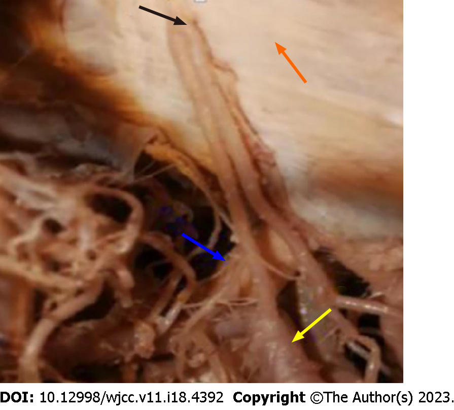
Figure 1 Perforating artery (black arrow) originated from anterior superior wall of A1 segment of anterior cerebral artery (yellow arrow), with the dura mater (orange arrow) moved forward.
The small branches and the dura mater traffic were folded back into the precribrum. Blue arrow indicates the optic nerve on the same side.
Figure 2 A posterior circulation superior cerebellar artery aneurysm with variant origin of the ophthalmic artery was present on digital subtraction angiography.
The lateral (A) and positive (B) position of the left internal carotid artery angiography showed two blood vessels at the end of the internal carotid artery. One was the thinner anterior cerebral artery (blue arrow) and the other was the thicker middle cerebral artery (cyan arrow). The abnormal origin of ophthalmic artery (yellow arrow) was on the A1 segment of the anterior cerebral artery. The orange arrow indicates the embryonic posterior cerebral artery. The normal origin of the ophthalmic artery should be on the ophthalmic artery segment (black arrow); C: shows angiography of the right internal carotid artery with evidence of a similar structure to the left.
- Citation: Mo ZX, Li W, Wang DF. Perforating and ophthalmic artery variants from the anterior cerebral artery: Two case reports. World J Clin Cases 2023; 11(18): 4392-4396
- URL: https://www.wjgnet.com/2307-8960/full/v11/i18/4392.htm
- DOI: https://dx.doi.org/10.12998/wjcc.v11.i18.4392


