Copyright
©The Author(s) 2023.
World J Clin Cases. Jun 26, 2023; 11(18): 4360-4367
Published online Jun 26, 2023. doi: 10.12998/wjcc.v11.i18.4360
Published online Jun 26, 2023. doi: 10.12998/wjcc.v11.i18.4360
Figure 1 Ultrasound characteristics of the left thyroid nodule.
A: Sonogram of the thyroid gland showing an irregular hypoechoic nodule (arrow) in the left thyroid; B: The nodule was near the trachea; C: Color Doppler view showing that the nodule had rich blood supply.
Figure 2 Cellular morphology of thyroid tissue aspired by fine needle.
A: Photomicrographs show the characteristics of cellular morphology of malignancy (hematoxylin-eosin stain, original magnification × 40); B: Hematoxylin-eosin stain, original magnification × 100; C: Hematoxylin-eosin stain, original magnification × 200; D: Hematoxylin-eosin stain, original magnification × 400.
Figure 3 Microphotographs of the thyroid metastasis.
A: Hematoxylin-eosin stain of the thyroid gland detects adenocarcinoma with mucinous features, consistent with metastatic rectal adenocarcinoma (original magnification × 40); B: Original magnification × 100; C: Original magnification × 200; D: Original magnification × 400.
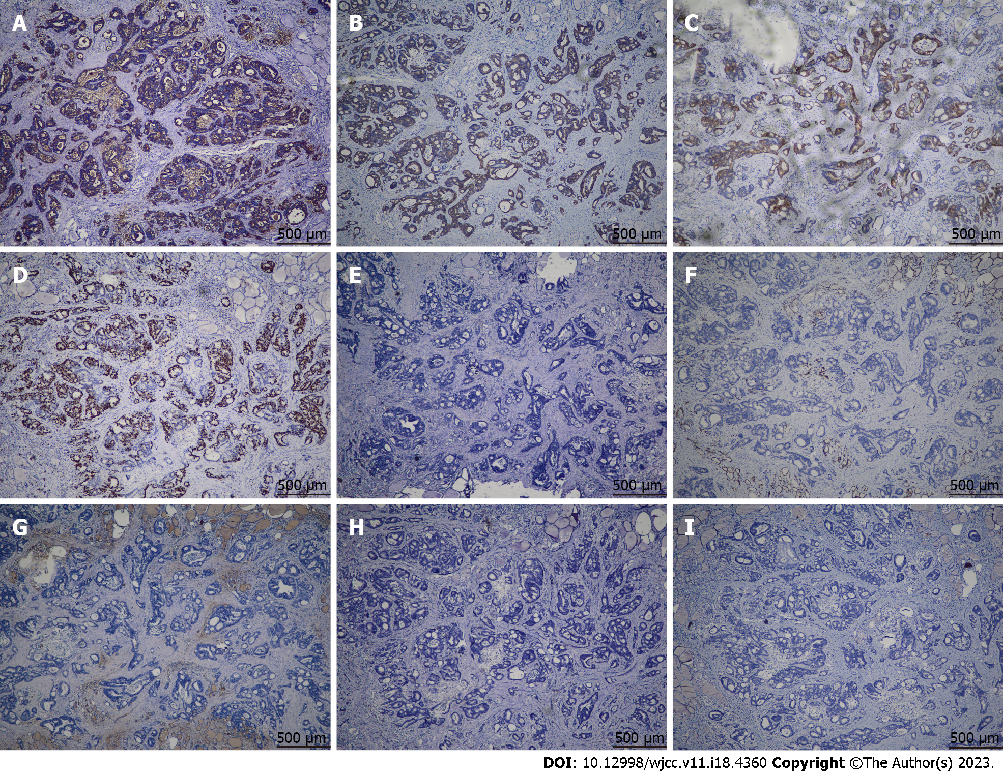
Figure 4 Immunohistochemical images showing metastatic carcinoma in the resected thyroid gland.
A-D: Tumor cells are positive for carcinoembryonic antigen (A), cytokeratin 20 (B), EMA (C), and Ki-67 (about 50%) (D); E-I: Tumor cells are negative for HBME1 (E), cytokeratin 7 (F), thyroglobulin (G), synaptophysin (H), and calcitonin (I).
Figure 5 Imaging characteristics of the recurrent left neck mass.
A: Ultrasonography of the neck showing a 2.50 cm × 1.64 cm × 1.17 cm mass within left strap muscles and sternocleidomastoid muscle (white arrows; sagittal view); B: Color Doppler view showing the mass has rich blood supply; C: Computed tomography of the neck showing a 2.5 cm × 1.6 cm × 1.1 cm mass within left strap muscles and sternocleidomastoid muscle (white arrow; sagittal view); D: Contrast-enhanced scan showing that the mass was above the internal jugular vein (IJV), with the IJV depressed. IJV: Internal jugular vein; CCA: Common carotid artery.
- Citation: Chen Y, Kang QS, Zheng Y, Li FB. Solitary thyroid gland metastasis from rectal cancer: A case report and review of the literature. World J Clin Cases 2023; 11(18): 4360-4367
- URL: https://www.wjgnet.com/2307-8960/full/v11/i18/4360.htm
- DOI: https://dx.doi.org/10.12998/wjcc.v11.i18.4360


