Copyright
©The Author(s) 2023.
World J Clin Cases. Jun 26, 2023; 11(18): 4334-4340
Published online Jun 26, 2023. doi: 10.12998/wjcc.v11.i18.4334
Published online Jun 26, 2023. doi: 10.12998/wjcc.v11.i18.4334
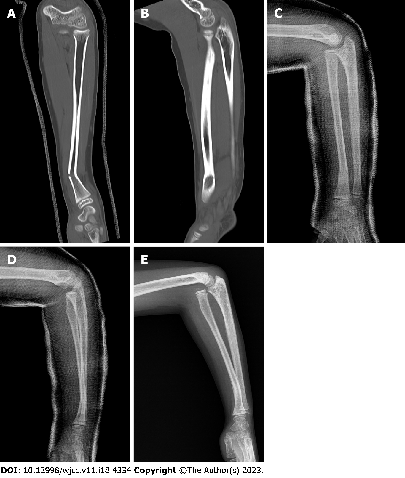
Figure 1 Images.
A and B: Computed tomography images of the left forearm performed at the time of injury. A linear fracture with angulation is seen in the distal third of the radius (A); there is no radial head dislocation in the elbow joint (B); C and D: Radiographs of the left forearm performed two weeks after the injury. The radial fracture is well reduced and shows no evidence of displacement progression (C); a mild subluxation of the radial head, which was not observed at the time of injury, is seen at the elbow joint (D); E: Preoperative radiographs of the left forearm performed three months after the injury. Malunion with a posterior convex deformity of 22° at the radial fracture site associated with complete anterior dislocation of the radial head is shown.
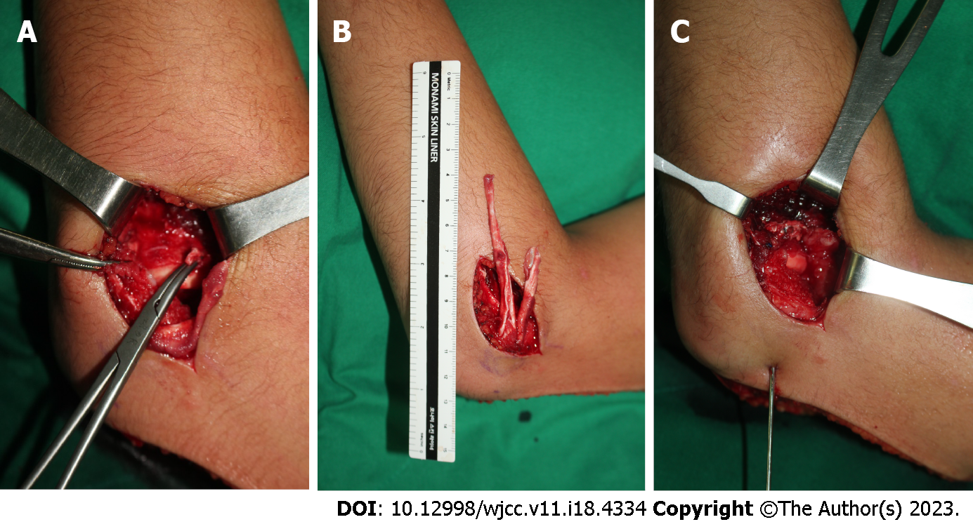
Figure 2 Surgical findings of dislocation of the radial head associated with malunited radial shaft.
A: The elbow joint was approached from the lateral aspect. Soft tissue, such as the joint capsule, was impinged between the capitellum, and the radial head and annular ligament were ruptured, making it difficult to recognize; B: An 8 cm × 1 cm strip of lateral triceps fascia and another 5 cm × 1 cm strip were harvested using a posterior incision, leaving the distal attached to the proximal ulna; C: The long fascial strip was passed around the neck of the radius from behind to forward and sutured back to itself and the ulnar periosteum. The short strip was additionally augmented to the anterior part of the long strip. Finally, reduction of the radial head was held using transarticular Kirschner wires with the elbow in 90° of flexion and the forearm in neutral rotation.
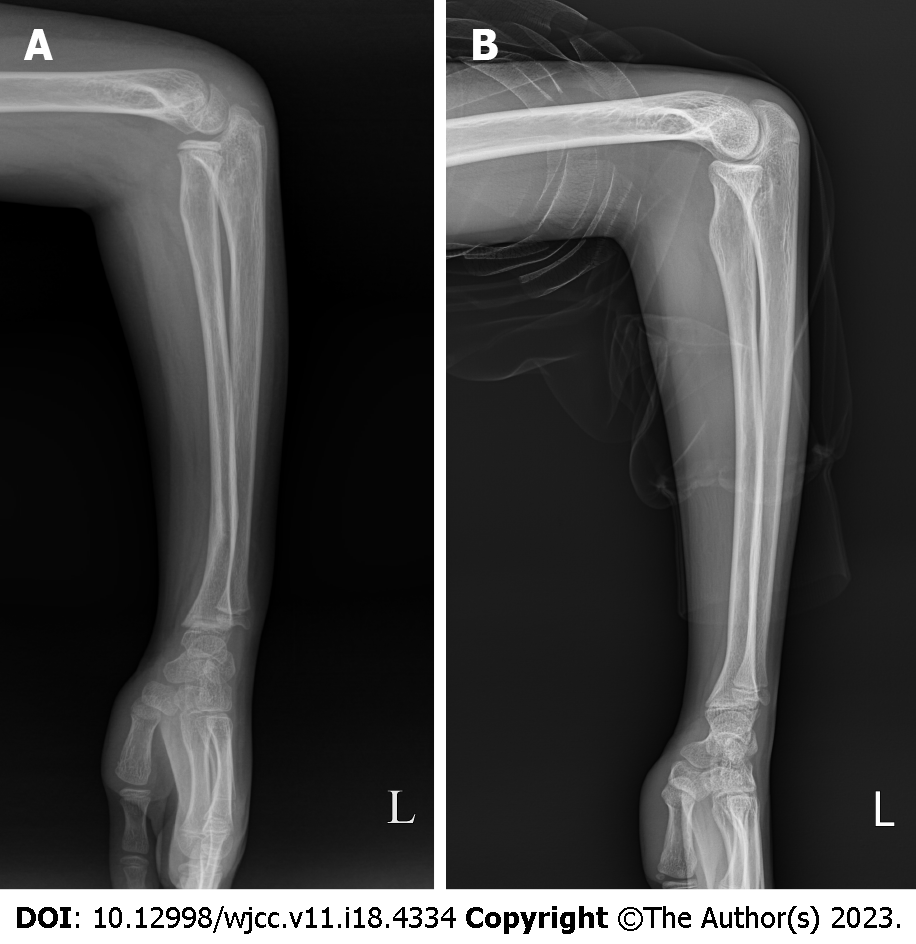
Figure 3 Radiographs of the left forearm performed during postoperative follow-up.
A: The radial head dislocation was well reduced within the radiocapitellar joint in the forearm lateral radiograph taken three months postoperatively, although angulation of the distal radius remained; B: Four years after surgery, the angulation deformity of the distal radius was corrected with the restoration of the normal curvature of the radius, showing no recurrence of radial head dislocation.
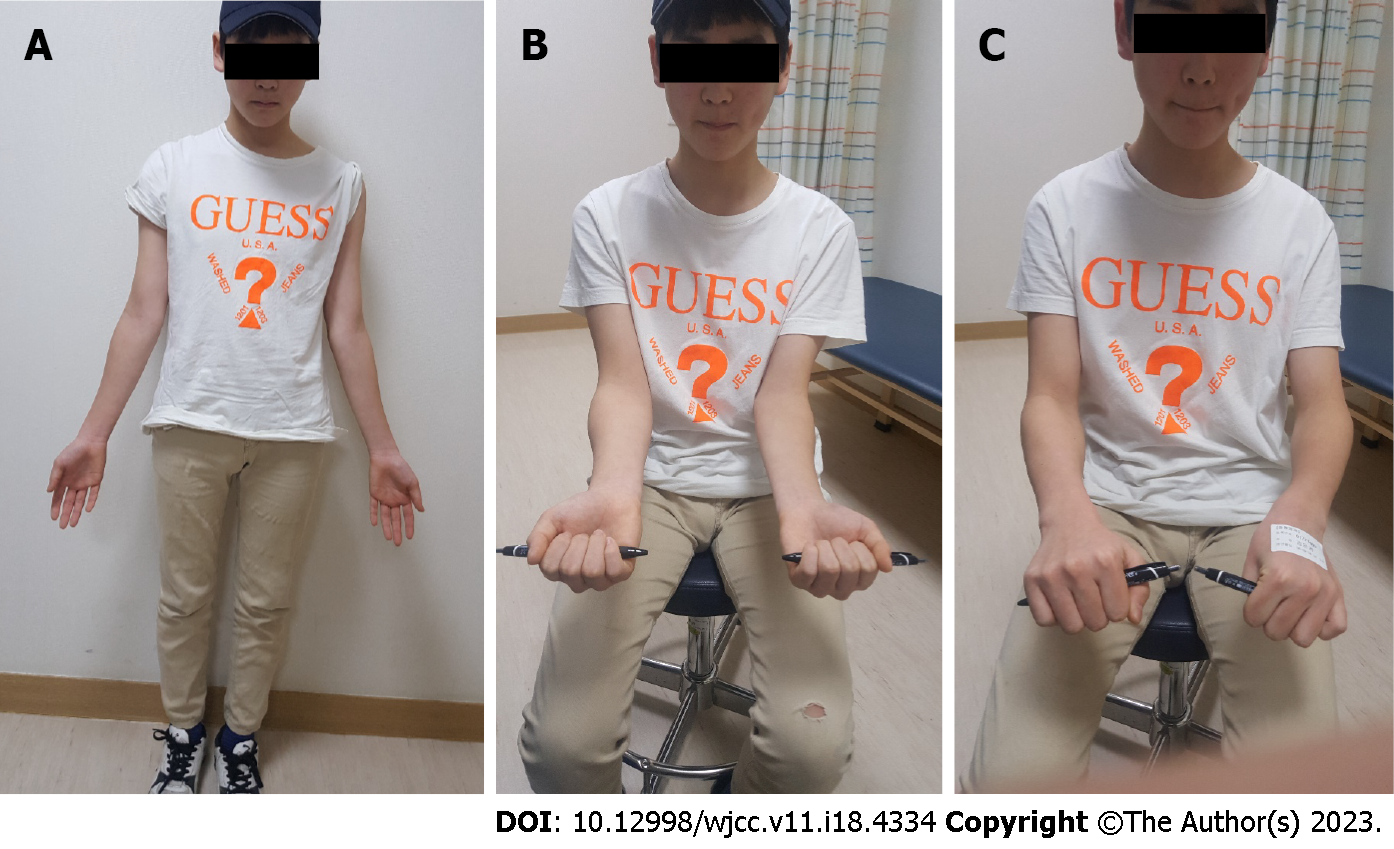
Figure 4 Clinical photographs taken at the last follow-up.
A: The active range of motion was 130° of flexion, 0° of extension; B: 85° of supination; C: 80° of pronation for the left elbow in the last follow-up photographs.
- Citation: Kim KB, Wang SI. Delayed dislocation of the radial head associated with malunion of distal radial fracture: A case report. World J Clin Cases 2023; 11(18): 4334-4340
- URL: https://www.wjgnet.com/2307-8960/full/v11/i18/4334.htm
- DOI: https://dx.doi.org/10.12998/wjcc.v11.i18.4334


