Copyright
©The Author(s) 2023.
World J Clin Cases. Jun 26, 2023; 11(18): 4313-4317
Published online Jun 26, 2023. doi: 10.12998/wjcc.v11.i18.4313
Published online Jun 26, 2023. doi: 10.12998/wjcc.v11.i18.4313
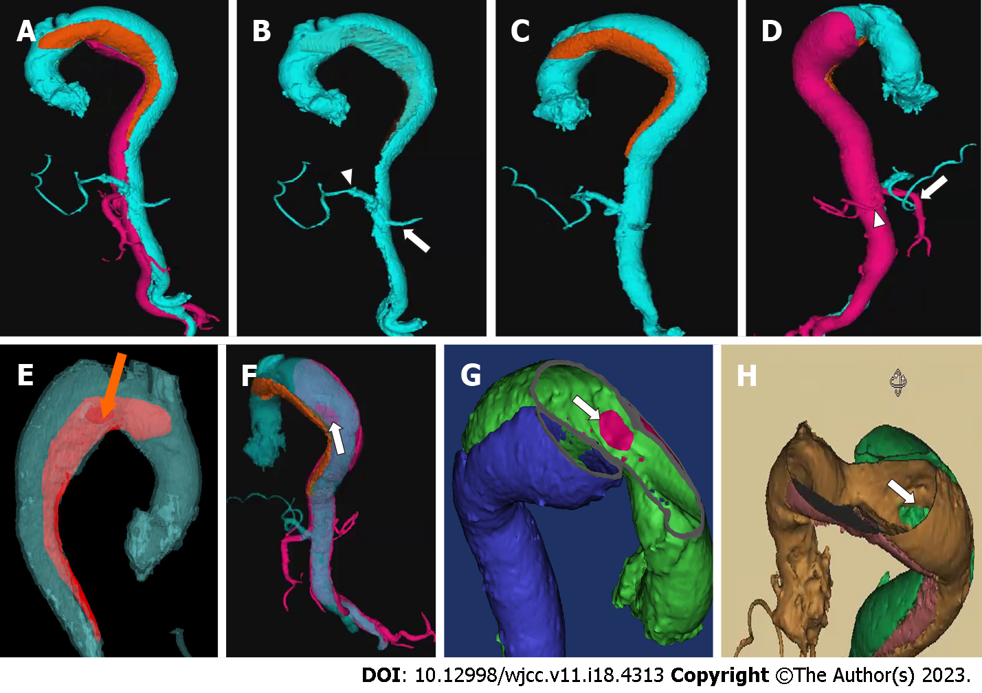
Figure 1 Image of medical 3D printing and modeling system for operative project planning.
A: Three channels with different colors were clearly shown in this case by a 3D modeling image; B: The true lumen depicted in blue color was collapsed with the celiac artery (white arrowhead) and the left renal artery (white arrow) originated from it; C: One of the false lumen (in orange) started from the less curvature of the aortic arch, whose entry tear was at the front wall of the aorta; D: The other false lumen (in pink) started from the greater curvature of the distal aortic arch and extended to the right common iliac artery, involving the superior mesentery artery (white arrow) and the right renal artery (white arrowhead). The second entry (to pink false lumen) was located at the back wall of the aorta; E: The entry tear of false lumen (in red) started from the less curvature of the aortic arch could be clearly recognized at the front wall of the aorta from outside (orange arrow); F: The second entry of the false lumen (in pink) started from the greater curvature of the distal aortic arch was located at the back wall of the aorta, as seen from the outside (white arrow); G: The entry tear of false lumen (in blue) started from the less curvature of the aortic arch could be clearly recognized within the true lumen (white arrow); H: The second entry of the false lumen (in green) started from the greater curvature of the distal aortic arch was located at the back wall of the aorta, as seen within the true lumen (white arrow).
Figure 2 Angiographic findings during the procedure.
A: Preoperative angiogram showed huge aneurysm caused by three-channeled aortic dissection; B: After stent graft deployment, the primary entries of both two false lumens were sealed without type I endoleaks as shown on the complete angiogram.
- Citation: Lu WF, Chen G, Wang LX. Morphological features and endovascular repair for type B multichanneled aortic dissection: A case report. World J Clin Cases 2023; 11(18): 4313-4317
- URL: https://www.wjgnet.com/2307-8960/full/v11/i18/4313.htm
- DOI: https://dx.doi.org/10.12998/wjcc.v11.i18.4313


