Copyright
©The Author(s) 2023.
World J Clin Cases. Jun 26, 2023; 11(18): 4295-4305
Published online Jun 26, 2023. doi: 10.12998/wjcc.v11.i18.4295
Published online Jun 26, 2023. doi: 10.12998/wjcc.v11.i18.4295
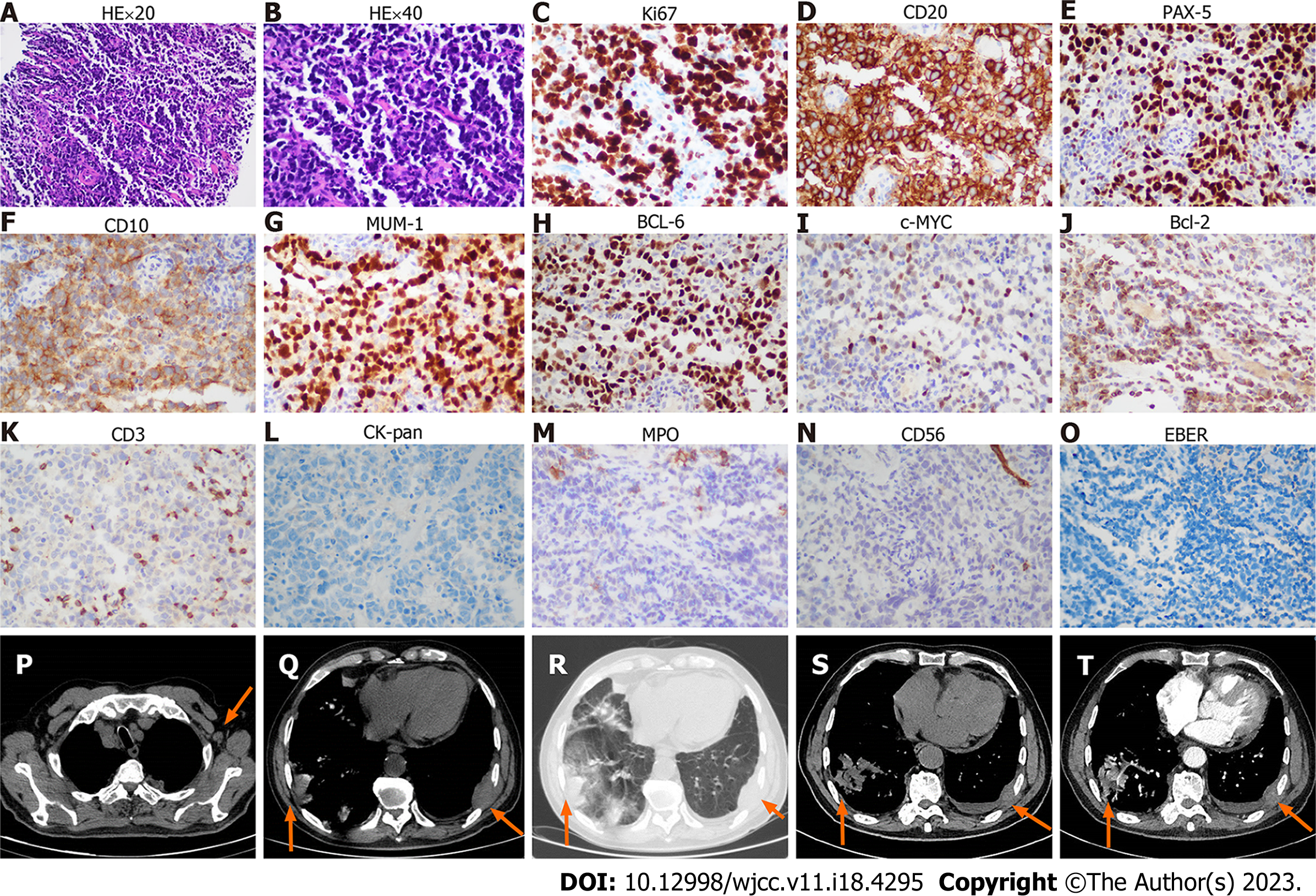
Figure 1 The biopsy and imaging of chest wall mass.
A-O: The HE and immunohistochemistry of chest wall mass; A: Hematoxylin-Eosin (HE) staining × 20 objective; B: HE staining × 40 objective; C-O: The immunohistochemistry shows Ki-67 (++, approximately 70%), CD20 (++), PAX-5 (++), CD10 (+), MUM-1 (++), BCL-6 (++), c-MYC (+, < 40%), BCL-2 (+, < 50%), CD3 (T cells few +), CK-pan (-), myeloperoxidase (MPO) (-), CD 56 (-), and EBER (-). P: Enlarged axillary lymph node (arrow) in the chest computed tomography (CT) scan; Q and R: Infiltration of masses (arrows) to the lung and chest wall on mediastinum window (Q) and lung window (R). S and T: Unenhanced (S) and contrast-enhanced (T) CT images shows the infiltration of masses (arrows) to the lung and chest wall on mediastinal window.
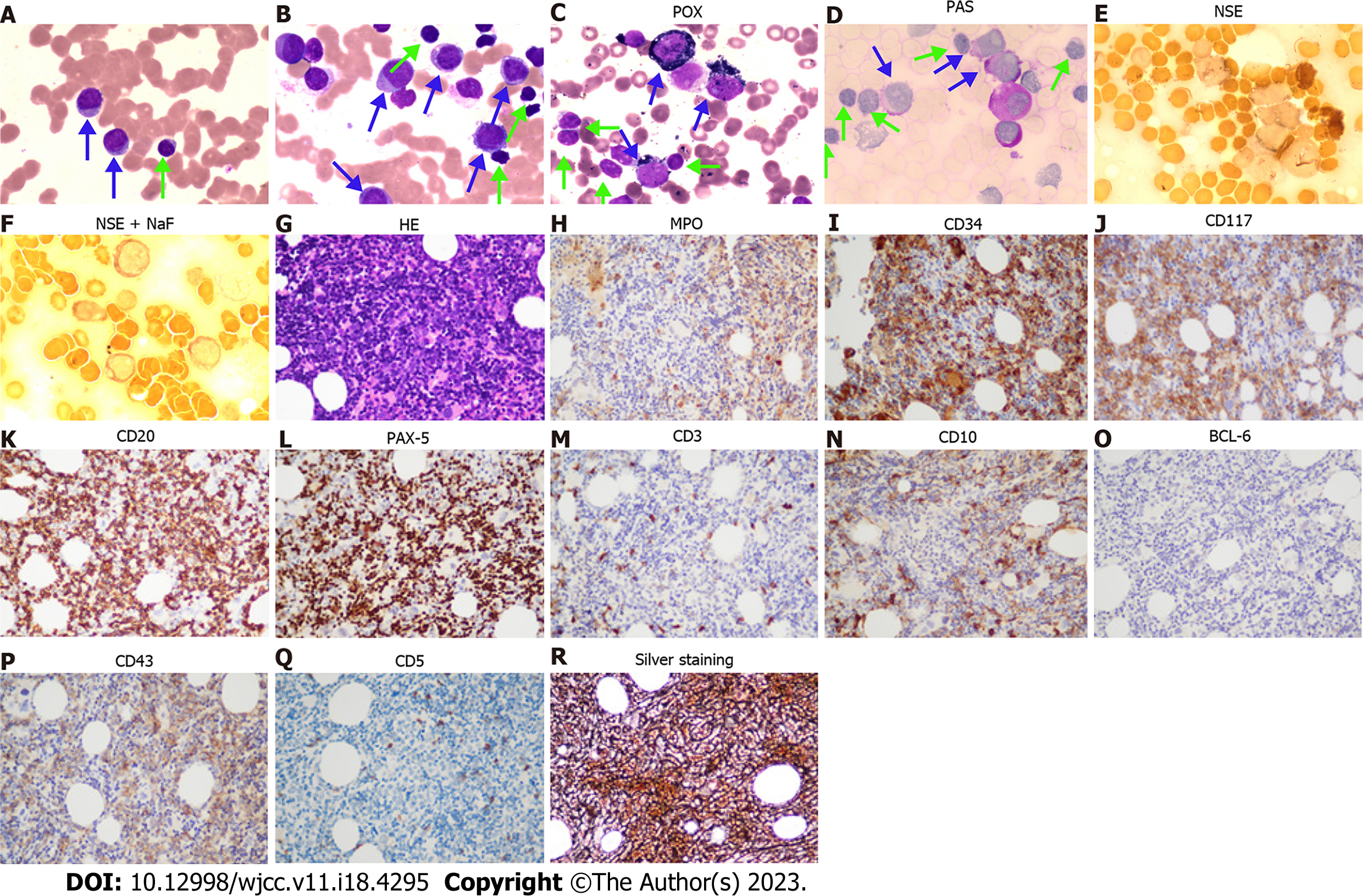
Figure 2 The cytomorphology of peripheral blood and bone marrow.
A: Peripheral blood smear (Wright staining, 100 × objective); B: Bone marrow smear (Wright staining, 100 × objective); C-F: Histochemistry staining for bone marrow smear. Peroxidase (52%, 99 points), periodic acid-Schiff stain (86%, 158 points), non-specific esterase (NSE) (32%, 35 points), and NSE + NaF (25%, 26 points) staining for bone marrow smear (Blue arrows direct myeloid blasts, while green arrows direct small lymphocytes); G-R: Bone marrow biopsy (40 × objective); G: Hematoxylin-Eosin staining; H-R: Immunohistochemistry shows myeloperoxidase (myelocytes +), CD34 (myelocytes ++), CD117 (myelocytes +), CD20 (B cells ++), PAX-5 (B cells ++), CD3 (T cells, few +), CD10 (few +), BCL-6(-), CD43(partial +), CD5 (T cells -), and silver staining (+++). POX: Peroxidase; PAS: Periodic acid-Schiff; NSE: Non-specific esterase; MPO: Myeloperoxidase.
Figure 3 Immunophenotype of bone marrow mononuclear cells.
Cells of group A (red color) are myeloid blasts expressing CD34, CD13, and CD117, and partially expressing CD33, HLA-DR, and myeloperoxidase (MPO). Cells of group B (blue color) are clonal B-lymphocytes expressing CD19, CD20, and Kappa, but no expressing CD103, CD25, CD11c, CD34, CD117, CD5, CD10, CD23, and Lambda. The results out the red box were from bone marrow sample obtained by bone marrow aspiration. The results in the red box were from peripheral blood gotten by venipuncture since the patient refused frequent bone marrow puncture.
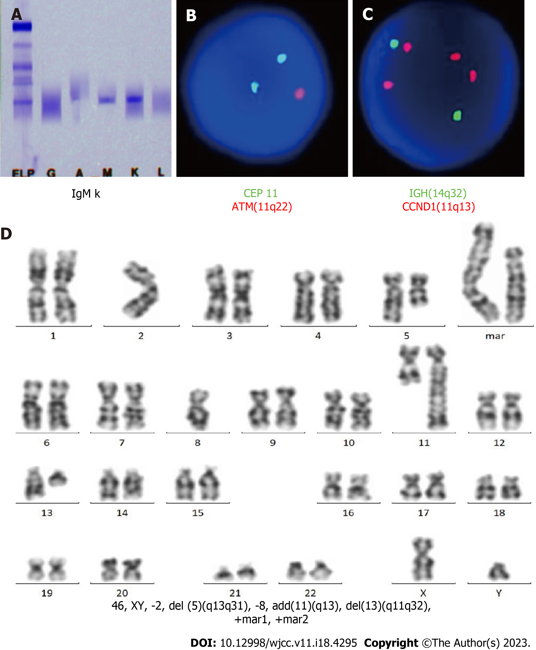
Figure 4 The immunofixation electrophoresis of serum and cytogenetic analysis of bone marrow mononuclear cells.
A: The monoclonal IgM Kappa is found by serum immunofixation electrophoresis; B and C: Fluorescence in situ hybridization of bone marrow mononuclear cells (BMMNCs) shows a deletion of ATM gene at 11q22 (2G1R 80%, 2G2R 17%, 1G1R 3%) and 4 copies of CCND1 gene at 11q13 (80%); D: The karyotype analysis of BMMNCs detects 46, XY, -2, del(5)(q13q31), -8, add(11)(q13), del(13)(q11q32), +mar1, +mar2 [5] or 46, XY, -2, del(5)(q13q31), -8, add(11)(q13), del(13)(q11q32), +mar1, +mar2 [20] under the stimulation of CpG-oligodeoxynucleotide.
- Citation: Zhang LB, Zhang L, Xin HL, Wang Y, Bao HY, Meng QQ, Jiang SY, Han X, Chen WR, Wang JN, Shi XF. Coexistence of diffuse large B-cell lymphoma, acute myeloid leukemia, and untreated lymphoplasmacytic lymphoma/waldenström macroglobulinemia in a same patient: A case report. World J Clin Cases 2023; 11(18): 4295-4305
- URL: https://www.wjgnet.com/2307-8960/full/v11/i18/4295.htm
- DOI: https://dx.doi.org/10.12998/wjcc.v11.i18.4295


