Copyright
©The Author(s) 2023.
World J Clin Cases. Jun 16, 2023; 11(17): 4159-4167
Published online Jun 16, 2023. doi: 10.12998/wjcc.v11.i17.4159
Published online Jun 16, 2023. doi: 10.12998/wjcc.v11.i17.4159
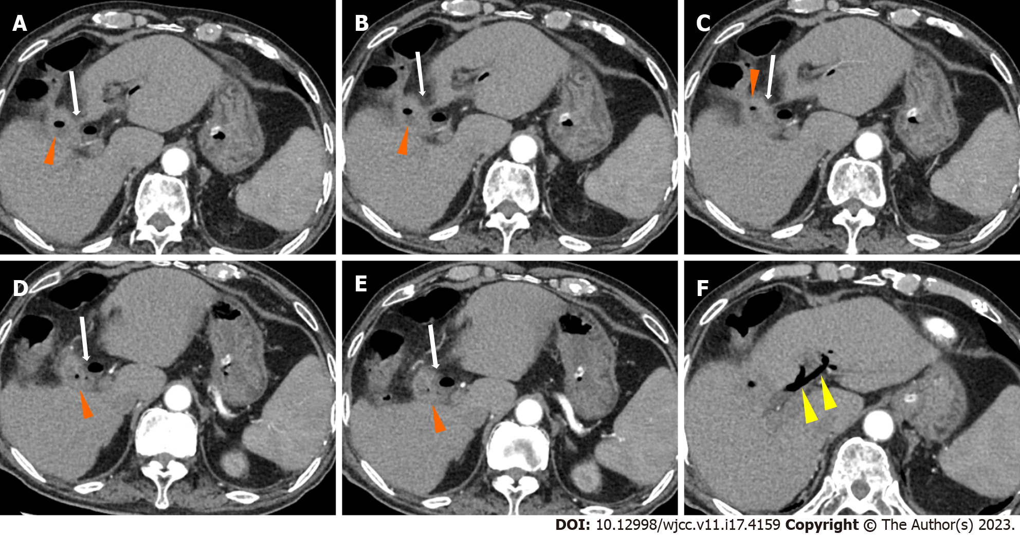
Figure 1 Abdominal contrast enhanced computed tomography (axial view).
A-E: Abdominal contrast computed tomography showed pneumatosis in the gallbladder (orange arrows), and the gallbladder communicated with the duodenal bulb (white arrows), indicating cholecystoduodenal fistula; F: Abdominal contrast computed tomography showed pneumobilia (yellow arrows).
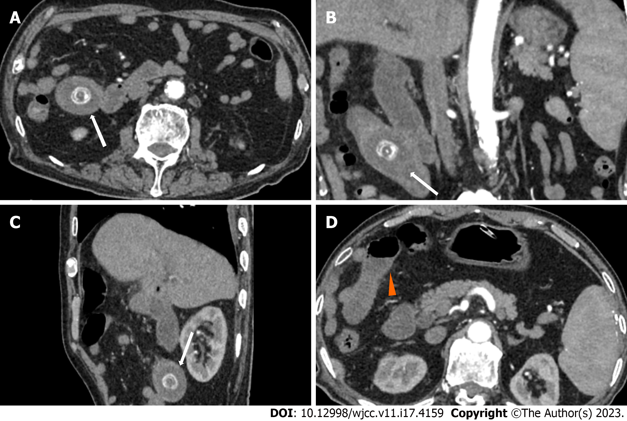
Figure 2 Abdominal contrast enhanced computed tomography (coronal view) showing cholecystoduodenal fistula.
A-D: Abdominal contrast computed tomography showed pneumatosis in the gallbladder (orange arrows), and the gallbladder communicated with the duodenal bulb (white arrows), indicating a cholecystoduodenal fistula.
Figure 3 Abdominal contrast enhanced computed tomography of the jejunal gallstone ileus.
A-C: A stone was shown in the upper jejunum (white arrows); D: Proximal intestinal effusion and dilatation (orange arrow).
Figure 4 Magnetic resonance cholangiopancreatography.
A-D: The gallbladder communicated with the duodenal bulb (white arrows), indicating cholecystoduodenal fistula; D: A short T2 signal as long as 25 mm in the upper jejunum with proximal intestinal dilatation was observed (orange arrow).
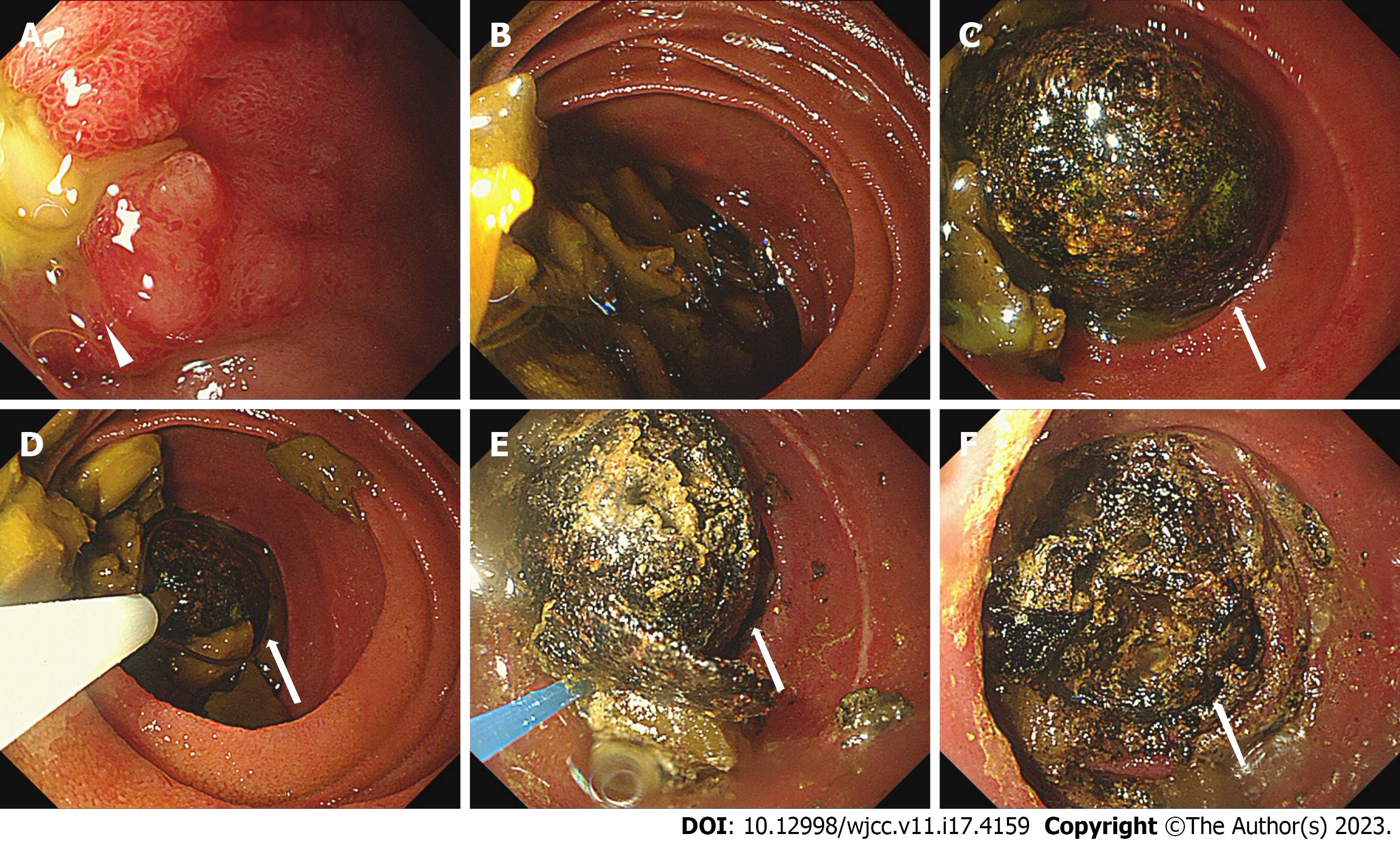
Figure 5 First propulsive enteroscopic examination.
A: We found a deep ulcer in the duodenal bulb close to the pylorus with yellow purulent secretion on the surface and mucosal edema (white arrow); B: Food residue was abundant proximal to the stone; C-F: We could see a stone incarceration in the upper jejunum 1.4 m from the incisor (white arrows); D: We failed to remove the stone with a snare; E-F: We performed laser lithotripsy and injected sodium bicarbonate on the stone.
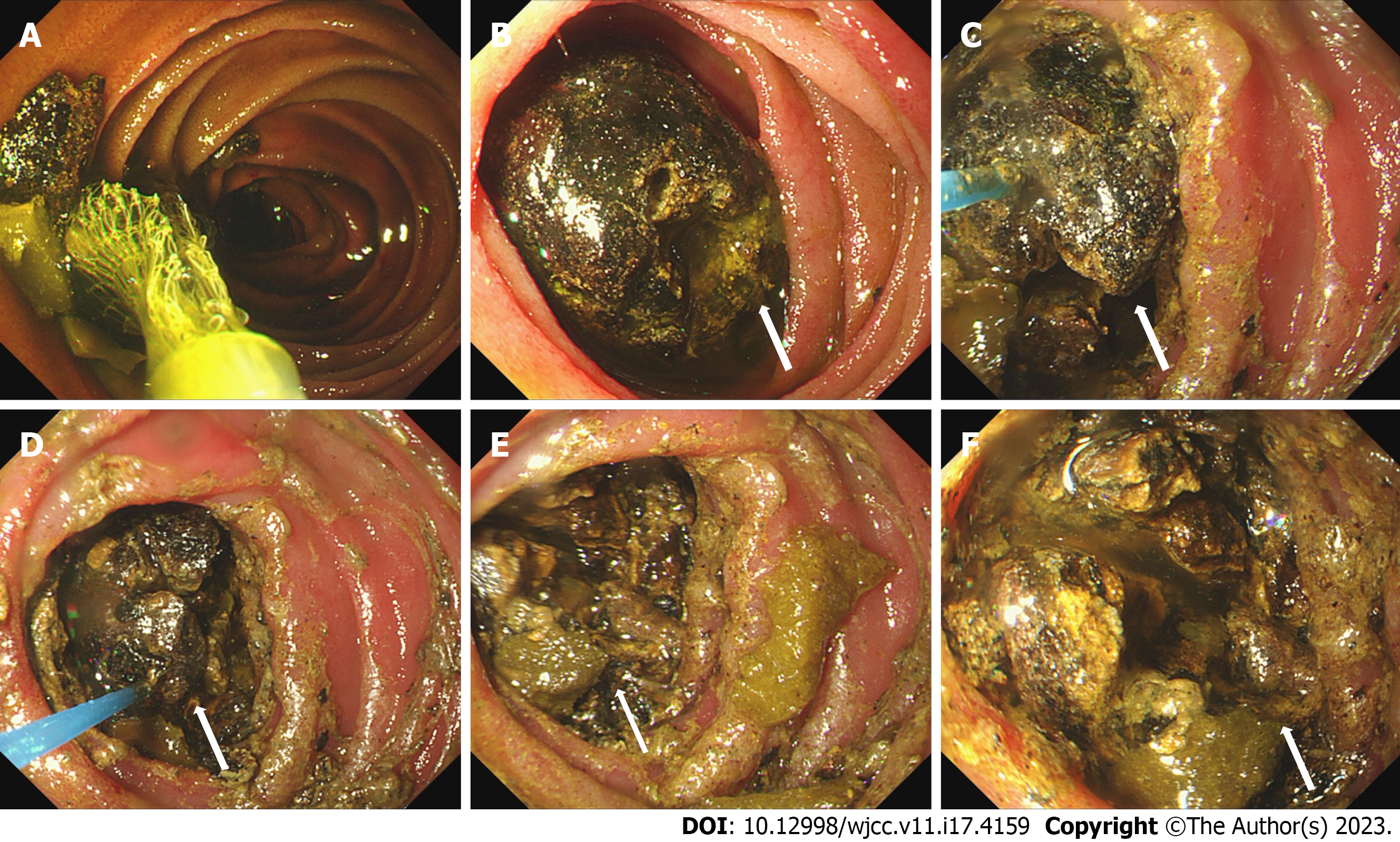
Figure 6 Second propulsive enteroscopic examination.
A: Food residue was abundant. We used a basket to remove the food residue; B: We could see a stone incarceration in the upper jejunum, which was smaller than before (white arrows); C-F: We performed laser lithotripsy and injected sodium bicarbonate on the stone.
- Citation: Fan WJ, Liu M, Feng XX. Endoscopic and surgical treatment of jejunal gallstone ileus caused by cholecystoduodenal fistula: A case report. World J Clin Cases 2023; 11(17): 4159-4167
- URL: https://www.wjgnet.com/2307-8960/full/v11/i17/4159.htm
- DOI: https://dx.doi.org/10.12998/wjcc.v11.i17.4159


