Copyright
©The Author(s) 2023.
World J Clin Cases. Jun 16, 2023; 11(17): 4123-4132
Published online Jun 16, 2023. doi: 10.12998/wjcc.v11.i17.4123
Published online Jun 16, 2023. doi: 10.12998/wjcc.v11.i17.4123
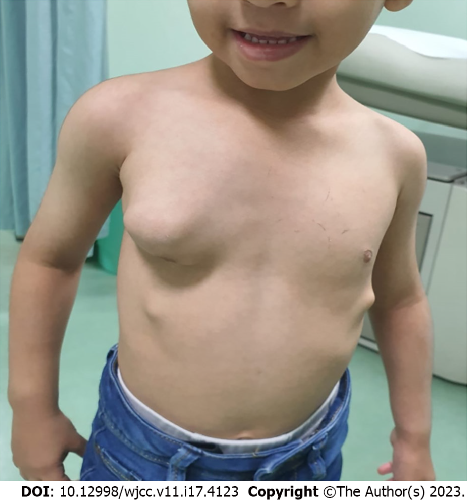
Figure 1 Image of the patient.
A large irregular right chest wall mass was observed. Several smaller lesions were obvious in the lower part of the chest bilaterally.
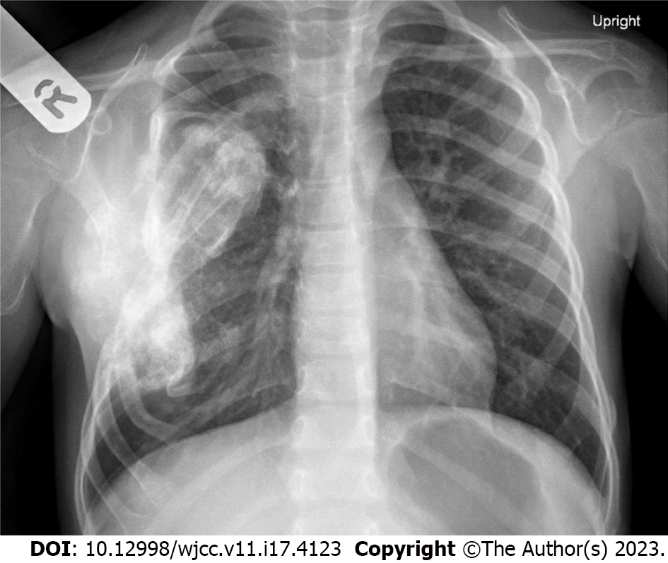
Figure 2 Chest X-ray of the child showing a large opacity over the right side of the chest.
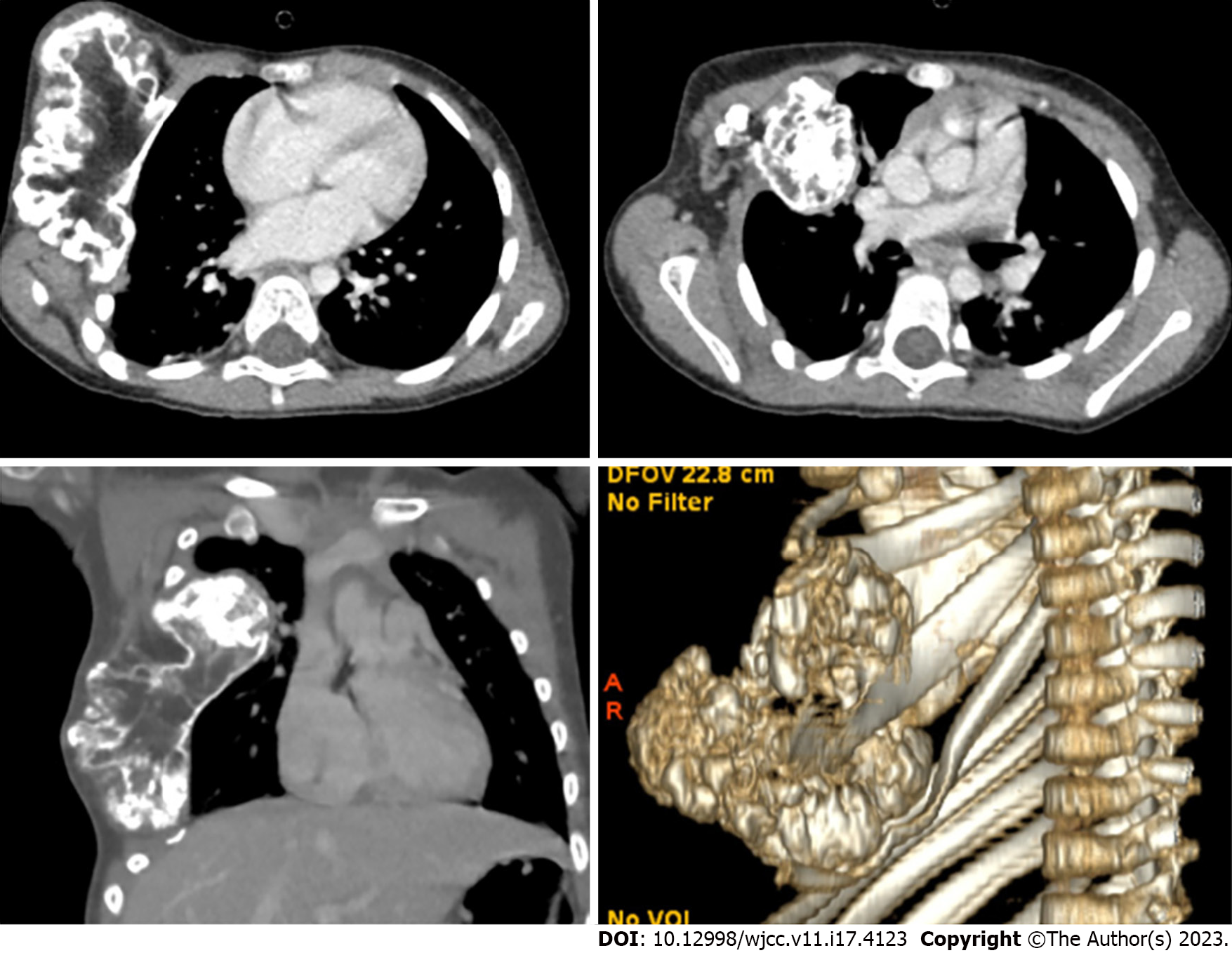
Figure 3 Computed tomography of the patient’s chest with three-dimensional reconstruction showed a large irregular bony chest wall lesion with an intrathoracic extension.
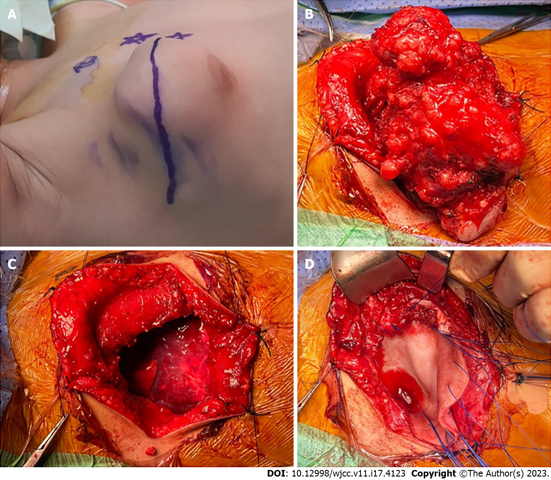
Figure 4 Intraoperative images of the resection and reconstruction.
A: Transverse upper thoracic incision over the lesion; B: The lesion exposed; C: The chest wall defect after the lesion was completely resected; D: Biological mesh was placed to cover the defect.
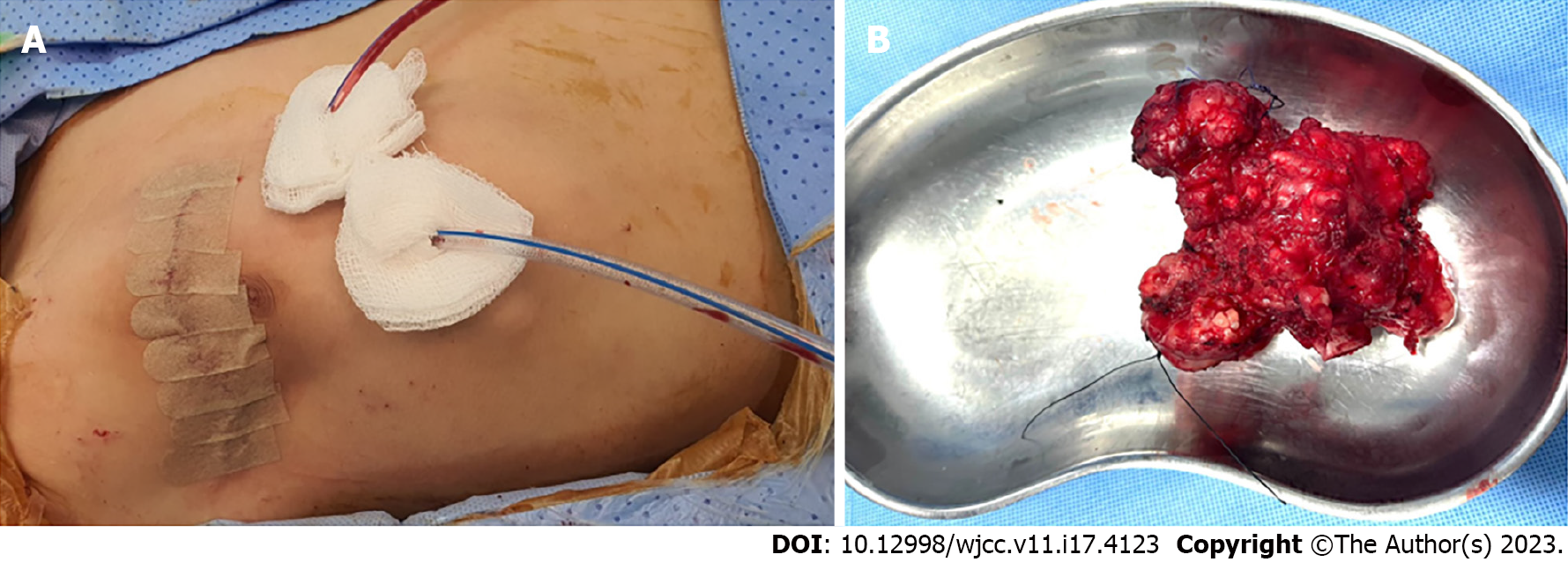
Figure 5 Postoperative images.
A: Two closed suction drains were placed after chest closure; B: The lesion after resection measured 10.5 cm × 9.0 cm × 5.5 cm.
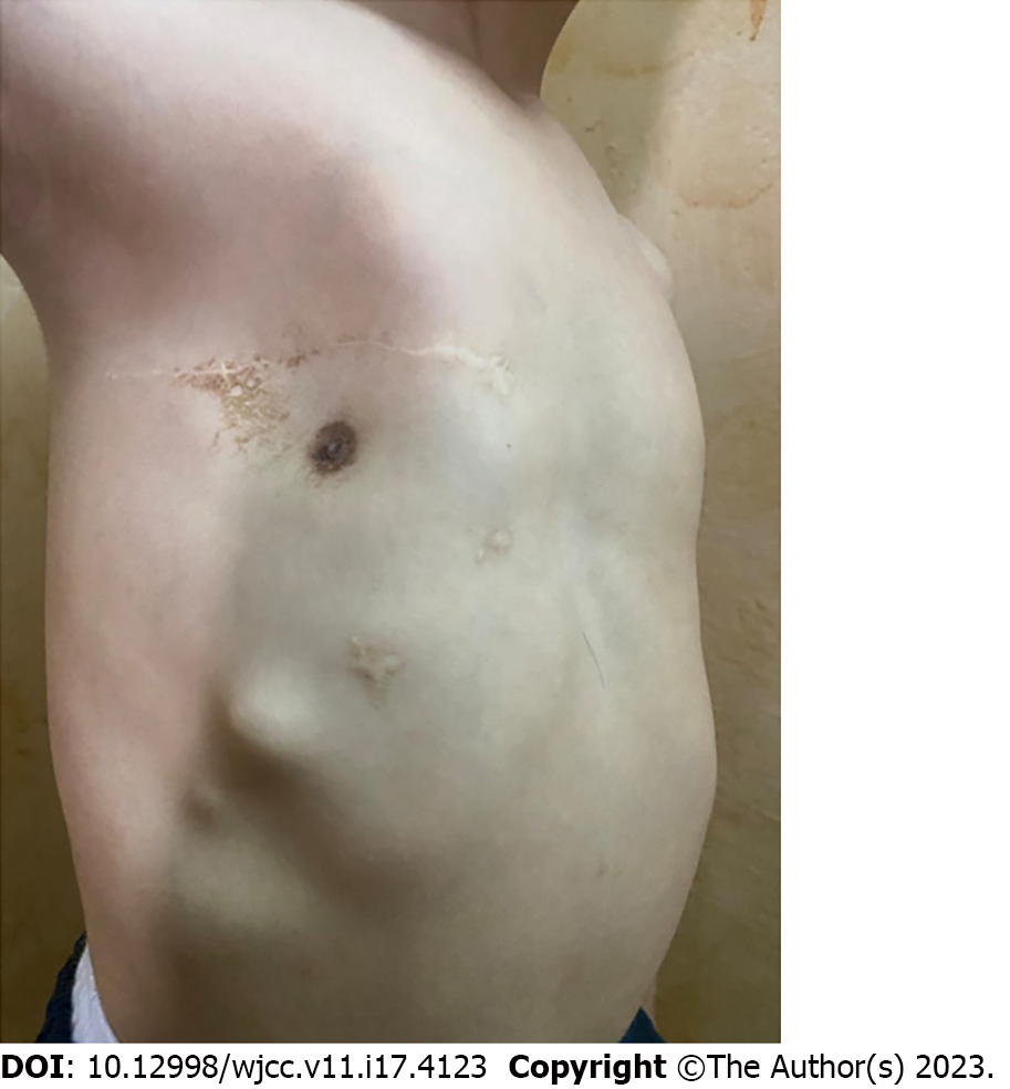
Figure 6 The patient’s chest wall 1 year after surgery with acceptable cosmesis.
- Citation: Alshehri A. Chest wall osteochondroma resection with biologic acellular bovine dermal mesh reconstruction in pediatric hereditary multiple exostoses: A case report and review of literature. World J Clin Cases 2023; 11(17): 4123-4132
- URL: https://www.wjgnet.com/2307-8960/full/v11/i17/4123.htm
- DOI: https://dx.doi.org/10.12998/wjcc.v11.i17.4123


