Copyright
©The Author(s) 2023.
World J Clin Cases. Jun 16, 2023; 11(17): 4098-4104
Published online Jun 16, 2023. doi: 10.12998/wjcc.v11.i17.4098
Published online Jun 16, 2023. doi: 10.12998/wjcc.v11.i17.4098
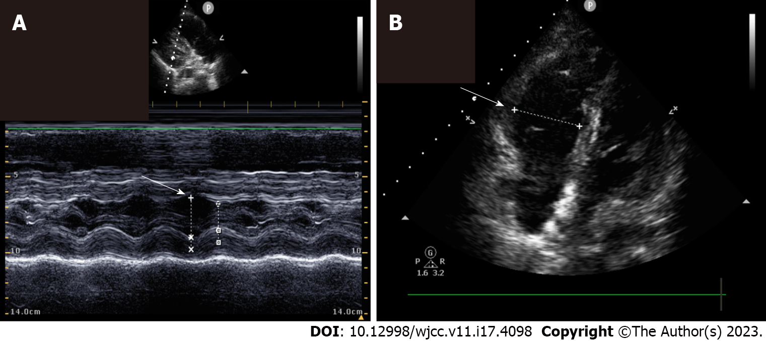
Figure 1 Echocardiographic examination of the patient in case 5.
A: Parasternal long axis view revealed that left ventricular end diastolic dimension was 26.2 mm; B: Apical 4 chamber view revealed that right ventricular end diastolic dimension was 36.1 mm. Right ventricle/left ventricle ratio > 1.
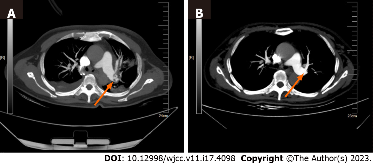
Figure 2 Pulmonary artery computed tomography angiography examination of the patient in case 1.
A: Emboli in the left main pulmonary artery (arrow); B: Emboli in the pulmonary artery were substantially decreased after 30 d of therapy (arrow).
- Citation: Qiu MS, Deng YJ, Yang X, Shao HQ. Cardiac arrest secondary to pulmonary embolism treated with extracorporeal cardiopulmonary resuscitation: Six case reports. World J Clin Cases 2023; 11(17): 4098-4104
- URL: https://www.wjgnet.com/2307-8960/full/v11/i17/4098.htm
- DOI: https://dx.doi.org/10.12998/wjcc.v11.i17.4098


