Copyright
©The Author(s) 2023.
World J Clin Cases. Jun 16, 2023; 11(17): 4090-4097
Published online Jun 16, 2023. doi: 10.12998/wjcc.v11.i17.4090
Published online Jun 16, 2023. doi: 10.12998/wjcc.v11.i17.4090
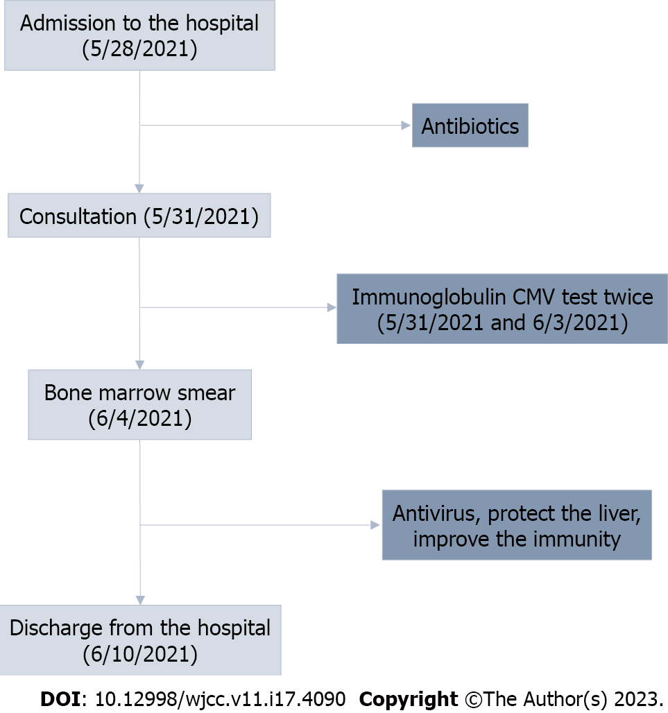
Figure 1 The process of treatment and diagnosis.
CMV: Cytomegalovirus.
Figure 2 Bone marrow smear.
2021.6.4: Visible phagocytes, phagocytic red blood cells, and platelets; granular hyperplasia is significantly active with left shift and dramatic changes.
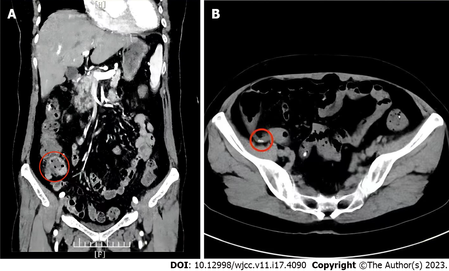
Figure 3 Abdominal contrast-enhanced computed tomography.
A: The local wall at the end of the ileum is thickened and the surrounding fat space is blurred with multiple small lymph nodes, suggestion: Inflammation may exist; B: Presence of appendiceal fecal stones.
- Citation: Sun FY, Ouyang BQ, Li XX, Zhang T, Feng WT, Han YG. Epstein-Barr virus-induced infection-associated hemophagocytic lymphohistiocytosis with acute liver injury: A case report. World J Clin Cases 2023; 11(17): 4090-4097
- URL: https://www.wjgnet.com/2307-8960/full/v11/i17/4090.htm
- DOI: https://dx.doi.org/10.12998/wjcc.v11.i17.4090


