Copyright
©The Author(s) 2023.
World J Clin Cases. Jun 16, 2023; 11(17): 4084-4089
Published online Jun 16, 2023. doi: 10.12998/wjcc.v11.i17.4084
Published online Jun 16, 2023. doi: 10.12998/wjcc.v11.i17.4084
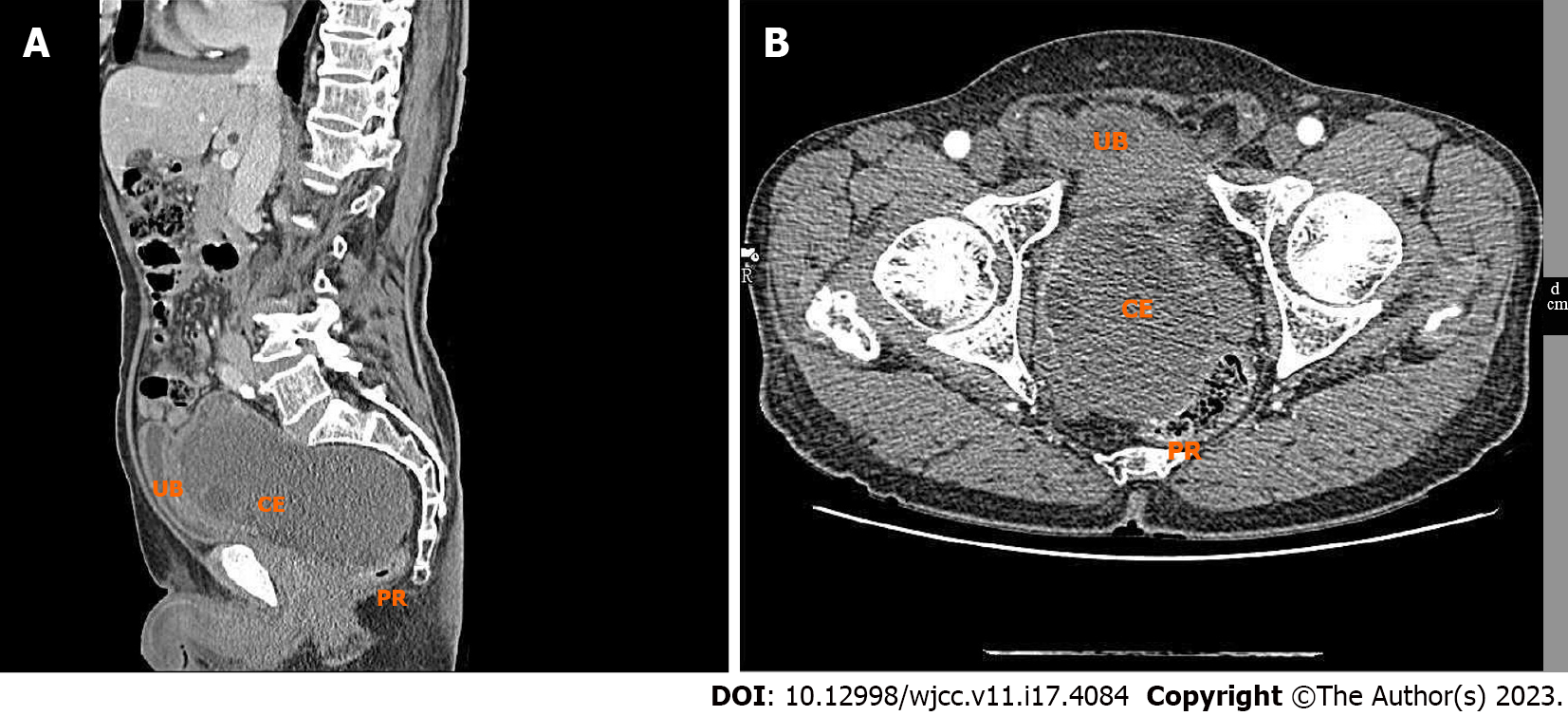
Figure 1 Computed tomography scans showed a space-occupying lesion with mixed density in the vesicorectal space, which was considered a benign lesion and was more likely to be hydatid disease.
A: The sagittal position of enhanced computed tomography (CT) of lesions; B: Enhanced CT coronal view of the lesion. UB: Urinary Bladder; CE: cystic echinococcosis; PR: Per Rectum.
Figure 2 Postoperative routine pathological evaluation showed cyst wall-like tissue with a thickness of 0.
2-1 cm, and appeared as gray-yellow with a slightly hard texture. Some gray-white materials were also observed with a soft texture. These findings indicate infection with Echinococcosis granulosus.
- Citation: Abulaiti Y, Kadi A, Tayier B, Tuergan T, Shalayiadang P, Abulizi A, Ahan A. Primary pelvic Echinococcus granulosus infection: A case report. World J Clin Cases 2023; 11(17): 4084-4089
- URL: https://www.wjgnet.com/2307-8960/full/v11/i17/4084.htm
- DOI: https://dx.doi.org/10.12998/wjcc.v11.i17.4084


