Copyright
©The Author(s) 2023.
World J Clin Cases. Jun 16, 2023; 11(17): 4072-4078
Published online Jun 16, 2023. doi: 10.12998/wjcc.v11.i17.4072
Published online Jun 16, 2023. doi: 10.12998/wjcc.v11.i17.4072
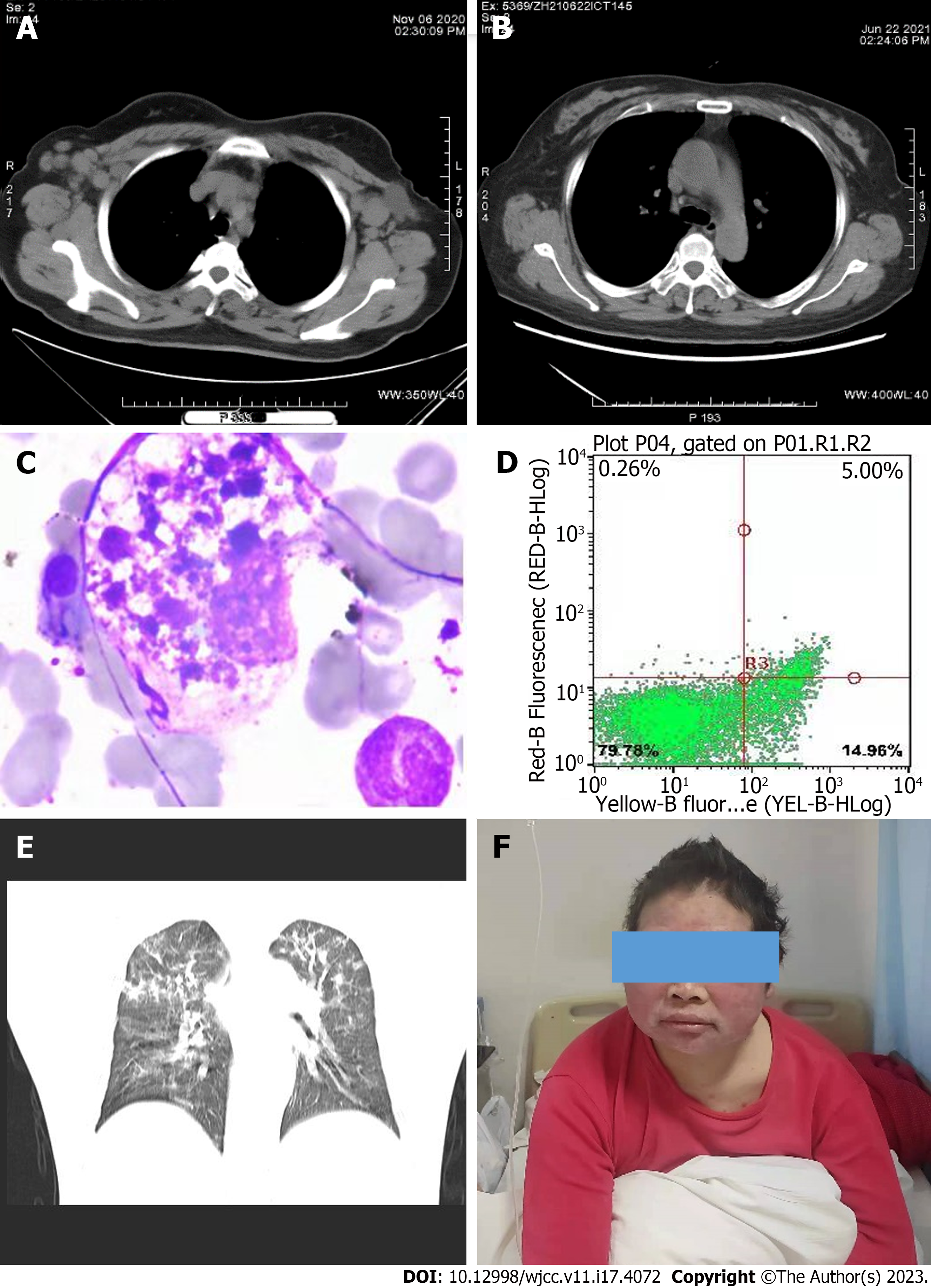
Figure 1 Laboratory finding and clinical manifestation in this patient before and after development of hemophagocytic lymphohisti
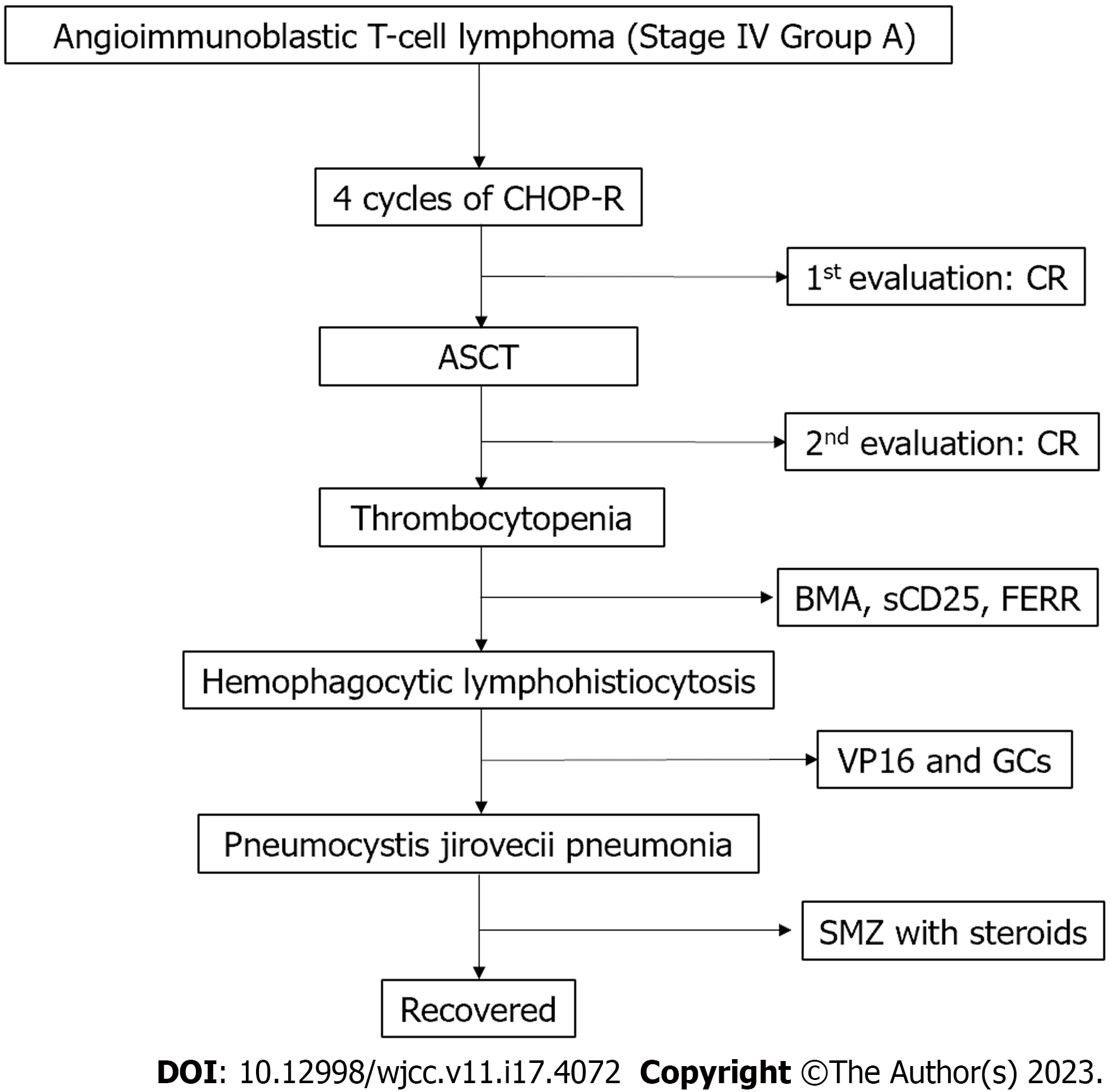
Figure 2 The flowchart of treatment process in this case.
CHOP-R: Cyclophosphamide, hydroxyl daunorubicin, vincristine, and prednisone; CR: Complete remission; ASCT: Autologous stem cell transplantation; GC: Glucocorticoids; FERR: Ferritin.
- Citation: Zhang ZR, Dou AX, Liu Y, Zhu HB, Jia HP, Kong QH, Sun LK, Qin AQ. Hemophagocytic lymphohistiocytosis after autologous stem cell transplantation in angioimmunoblastic T-cell lymphoma: A case report. World J Clin Cases 2023; 11(17): 4072-4078
- URL: https://www.wjgnet.com/2307-8960/full/v11/i17/4072.htm
- DOI: https://dx.doi.org/10.12998/wjcc.v11.i17.4072


