Copyright
©The Author(s) 2023.
World J Clin Cases. Jun 16, 2023; 11(17): 4065-4071
Published online Jun 16, 2023. doi: 10.12998/wjcc.v11.i17.4065
Published online Jun 16, 2023. doi: 10.12998/wjcc.v11.i17.4065
Figure 1 Transvaginal ultrasound of cesarian scar pregnancy.
A and B: White arrows indicate gestational matter (A) and dilated blood vessels (B) at the uterine scar.
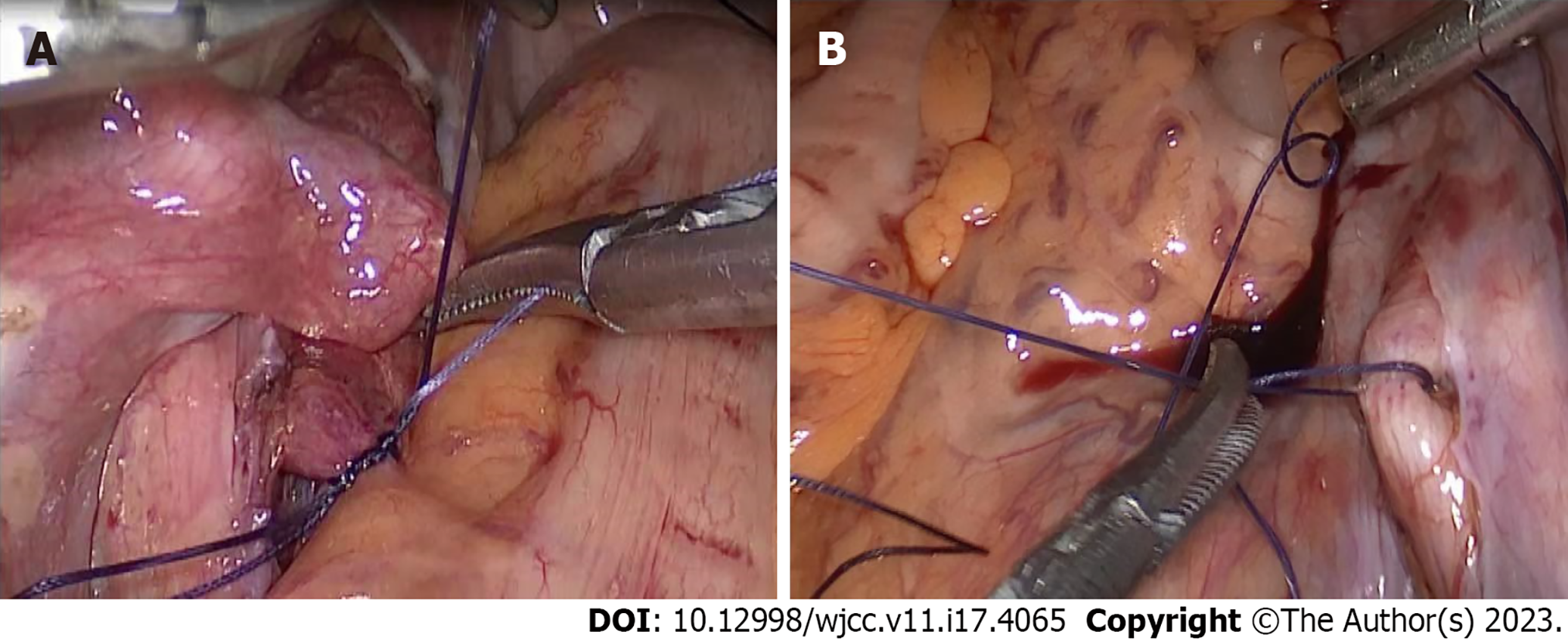
Figure 2 Intraoperative management of the internal iliac artery.
A: Temporary occlusion of the internal iliac artery; B: Release of the temporary occlusion of the internal iliac artery.
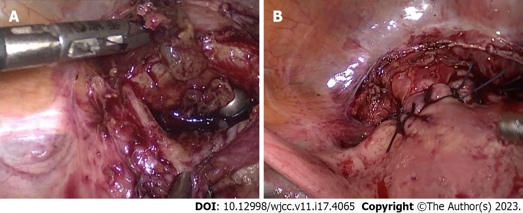
Figure 3 Pregnancy tissue removed and uterine scars repaired.
A: Removal of pregnancy tissue with spoon forceps through the vagina; B: Repair of the myometrium of the anterior uterine wall after scar trimming.
- Citation: Xie JP, Chen LL, Lv W, Li W, Fang H, Zhu G. Emergency internal iliac artery temporary occlusion after massive hemorrhage during surgery of cesarean scar pregnancy: A case report. World J Clin Cases 2023; 11(17): 4065-4071
- URL: https://www.wjgnet.com/2307-8960/full/v11/i17/4065.htm
- DOI: https://dx.doi.org/10.12998/wjcc.v11.i17.4065


