Copyright
©The Author(s) 2023.
World J Clin Cases. Jun 6, 2023; 11(16): 3907-3914
Published online Jun 6, 2023. doi: 10.12998/wjcc.v11.i16.3907
Published online Jun 6, 2023. doi: 10.12998/wjcc.v11.i16.3907
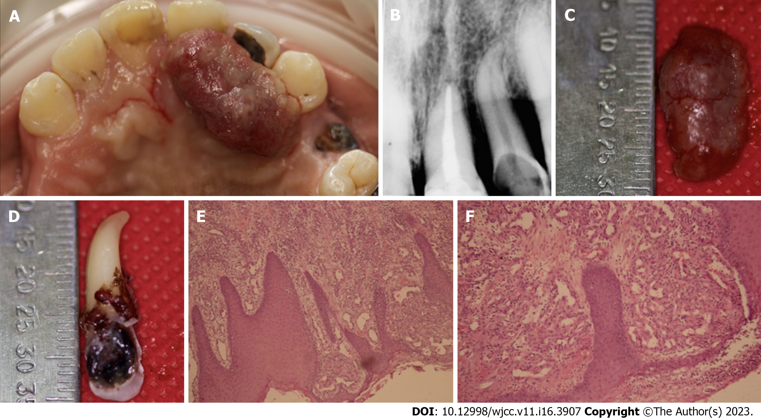
Figure 1 Clinical, radiographic and histological view of lesion case 1.
A: Exophytic and hemorrhagic lesion on the palate; B: Intraoral periapical radiograph of dental organ 22 showing alveolar crestal bone resorption; C: Excised specimen; D: Extracted dental organ 22 showing the extent of caries; E and F: Histopathological views.
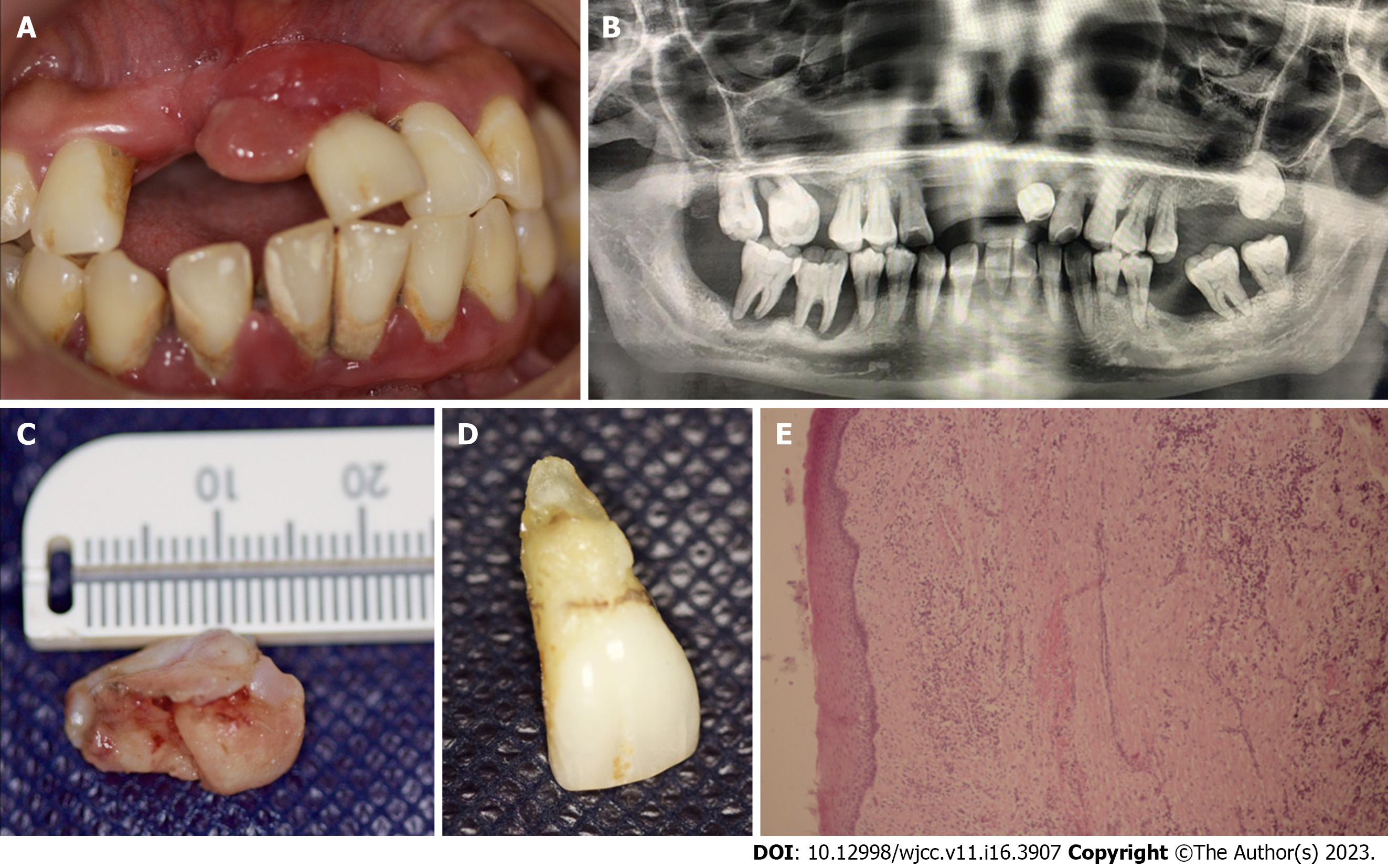
Figure 2 Clinical, radiographic and histological view of lesion case 2.
A: Exophytic lesion associated with dental organ 21; B: Panoramic radiography; C: Excised specimen; D: Extracted dental organ 21; E: Histopathological view.
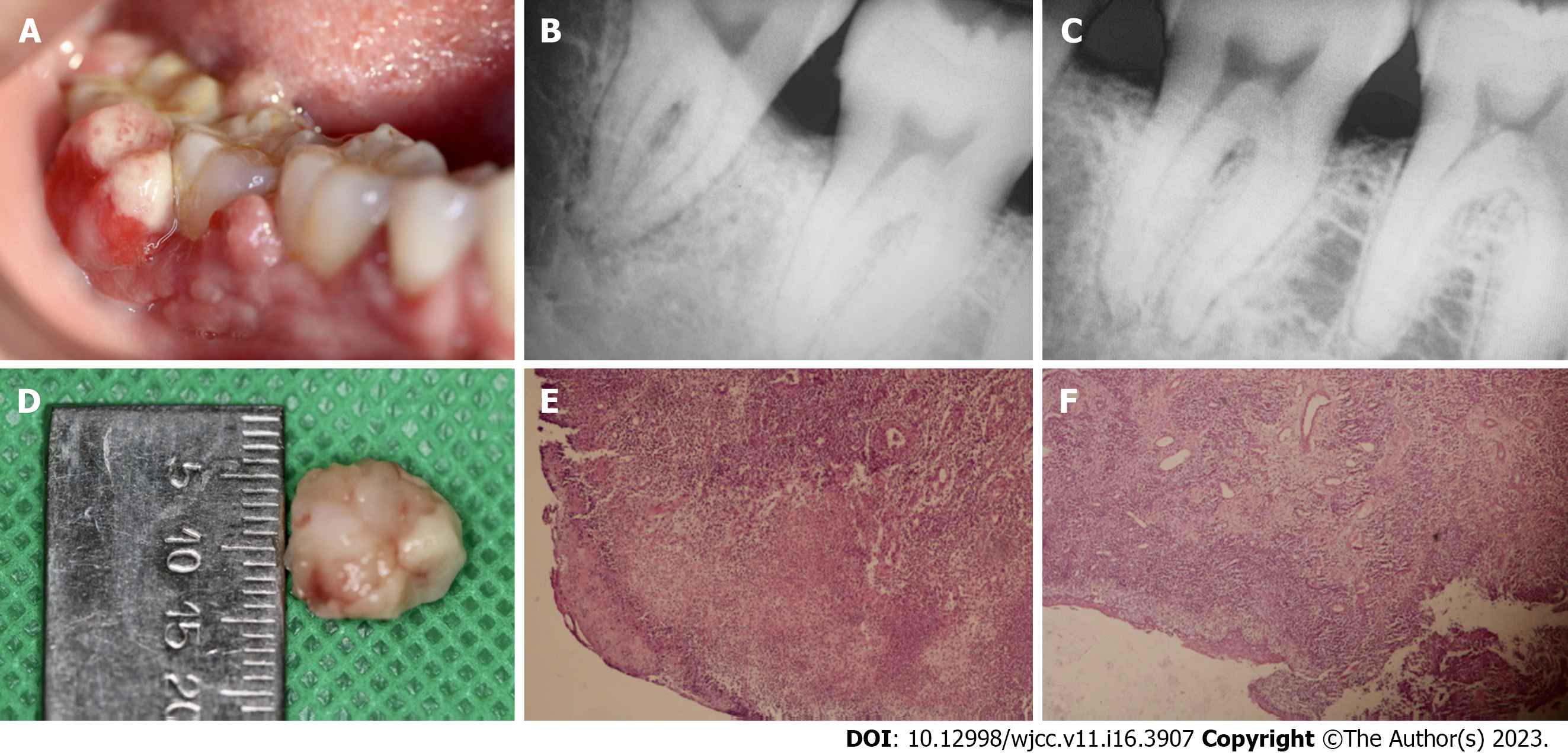
Figure 3 Clinical, radiographic and histological view of lesion case 3.
A: Exophytic lesion associated with dental organs 46 and 47; B and C: Intraoral periapical radiographs showing interproximal bone loss between teeth 47 and 48; D: Excised specimen; E and F: Histopathological views.
- Citation: Lomelí Martínez SM, Bocanegra Morando D, Mercado González AE, Gómez Sandoval JR. Unusual clinical presentation of oral pyogenic granuloma with severe alveolar bone loss: A case report and review of literature. World J Clin Cases 2023; 11(16): 3907-3914
- URL: https://www.wjgnet.com/2307-8960/full/v11/i16/3907.htm
- DOI: https://dx.doi.org/10.12998/wjcc.v11.i16.3907


