Copyright
©The Author(s) 2023.
World J Clin Cases. Jun 6, 2023; 11(16): 3899-3906
Published online Jun 6, 2023. doi: 10.12998/wjcc.v11.i16.3899
Published online Jun 6, 2023. doi: 10.12998/wjcc.v11.i16.3899
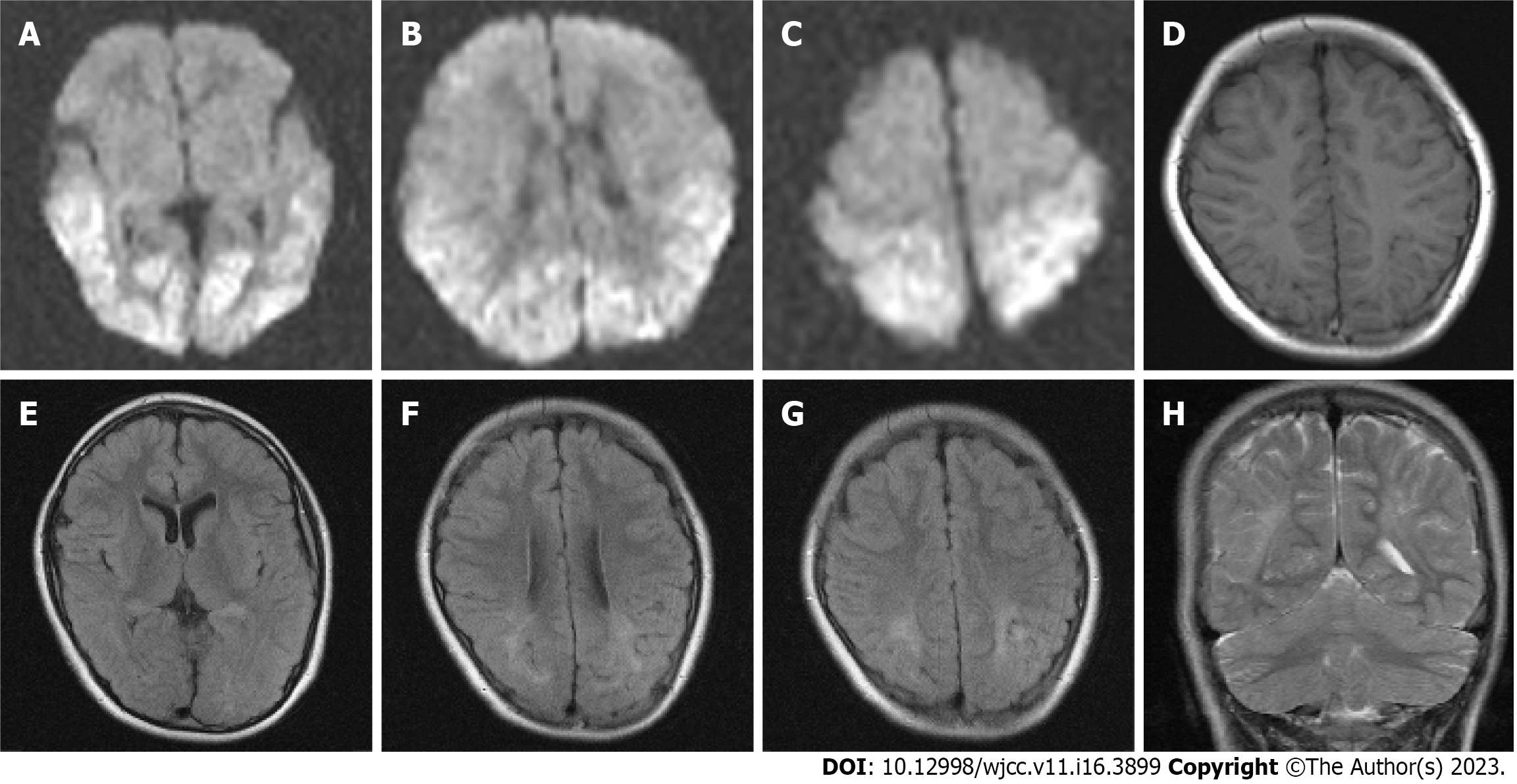
Figure 1 Diffusion-weighted images on day 4, and T1- and T2-weighted images, and fluid-attenuated inversion images at 8 years of age.
A-C: Diffusion-weighted images reveal cortical and subcortical hyperintensities in the bilateral occipital, parietal, and temporal lobes; D and F: T1-weighted image and fluid-attenuated inversion image show mild volume loss in the parieto-temporal region, compared with the frontal region, with minimal cortical changes; E-G: Fluid-attenuated inversion images show a high-intensity area in the white matter of the bilateral parieto-temporo-occipital lobes, which includes the periventricular region at the trigone of the lateral ventricles and centrum semiovale; H: T2-weighted image shows ulegyria in the bilateral parieto-temporal regions.
- Citation: Kurahashi N, Ogaya S, Maki Y, Nonobe N, Kumai S, Hosokawa Y, Ogawa C, Yamada K, Maruyama K, Miura K, Nakamura M. Reading impairment after neonatal hypoglycemia with parieto-temporo-occipital injury without cortical blindness: A case report. World J Clin Cases 2023; 11(16): 3899-3906
- URL: https://www.wjgnet.com/2307-8960/full/v11/i16/3899.htm
- DOI: https://dx.doi.org/10.12998/wjcc.v11.i16.3899


