Copyright
©The Author(s) 2023.
World J Clin Cases. Jun 6, 2023; 11(16): 3877-3884
Published online Jun 6, 2023. doi: 10.12998/wjcc.v11.i16.3877
Published online Jun 6, 2023. doi: 10.12998/wjcc.v11.i16.3877
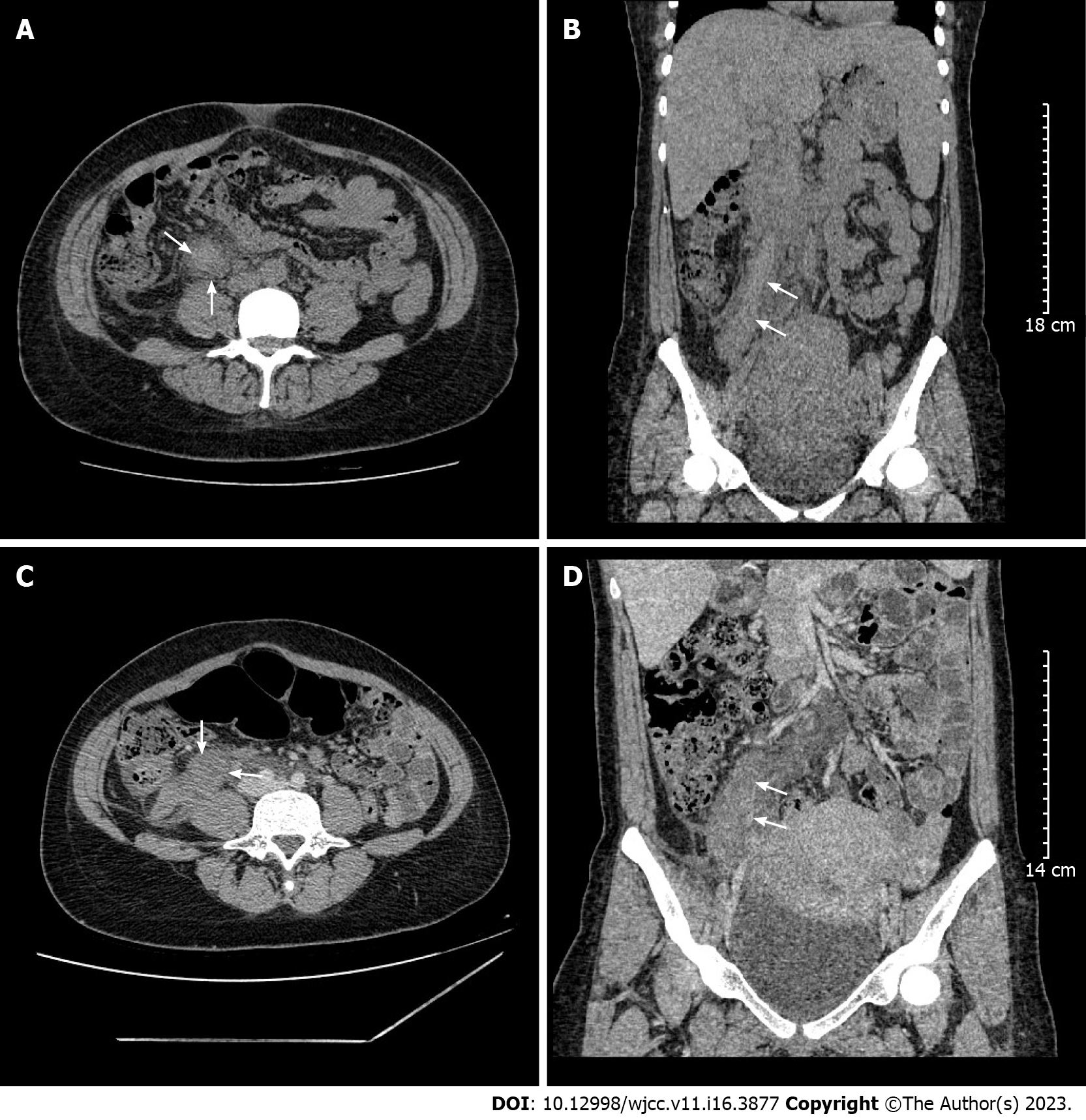
Figure 1 Computed tomography image showing right ovarian vein thrombosis in case 1 and case 2.
A: Axial view (arrows); B: Coronal view (arrows); C: Axial view (arrows); D: Coronal view (arrows).
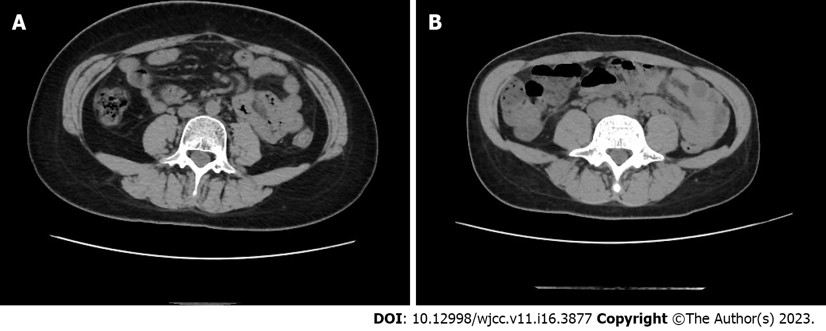
Figure 2 Computed tomography image.
A: The image of follow up computed tomography (CT) in case 1; B: The image of follow up CT in case 2.
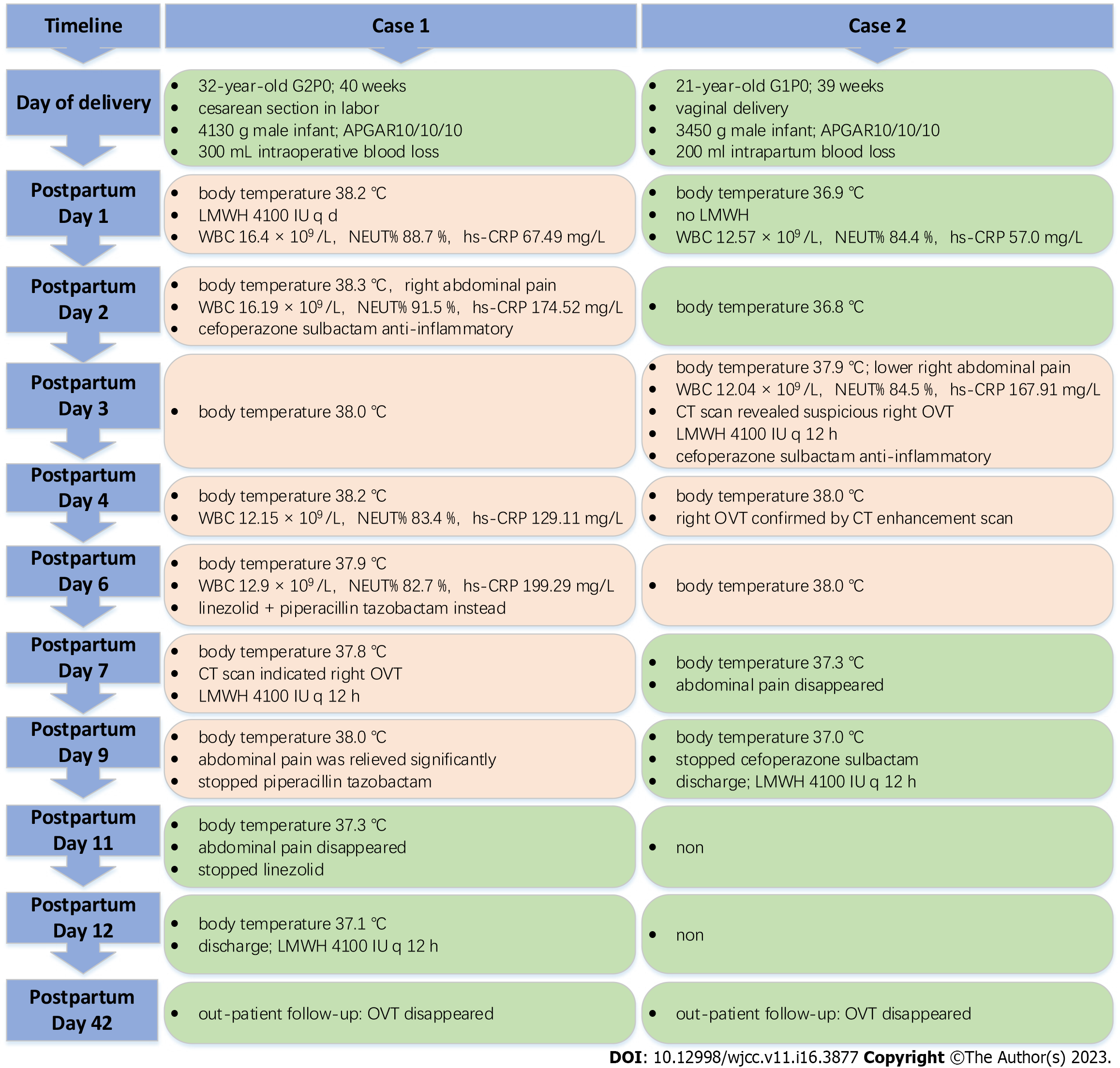
Figure 3 Time of events and findings.
LMWH: Low molecular weight heparin; WBC: White blood cell; NEUT%: Neutrophil percentage; hs-CRP: High sensitive C-reactive protein; CT: Computed tomography; OVT: Ovarian vein thrombosis.
- Citation: Zhu HD, Shen W, Wu HL, Sang X, Chen Y, Geng LS, Zhou T. Postpartum ovarian vein thrombosis after cesarean section and vaginal delivery: Two case reports. World J Clin Cases 2023; 11(16): 3877-3884
- URL: https://www.wjgnet.com/2307-8960/full/v11/i16/3877.htm
- DOI: https://dx.doi.org/10.12998/wjcc.v11.i16.3877


