Copyright
©The Author(s) 2023.
World J Clin Cases. Jun 6, 2023; 11(16): 3852-3857
Published online Jun 6, 2023. doi: 10.12998/wjcc.v11.i16.3852
Published online Jun 6, 2023. doi: 10.12998/wjcc.v11.i16.3852
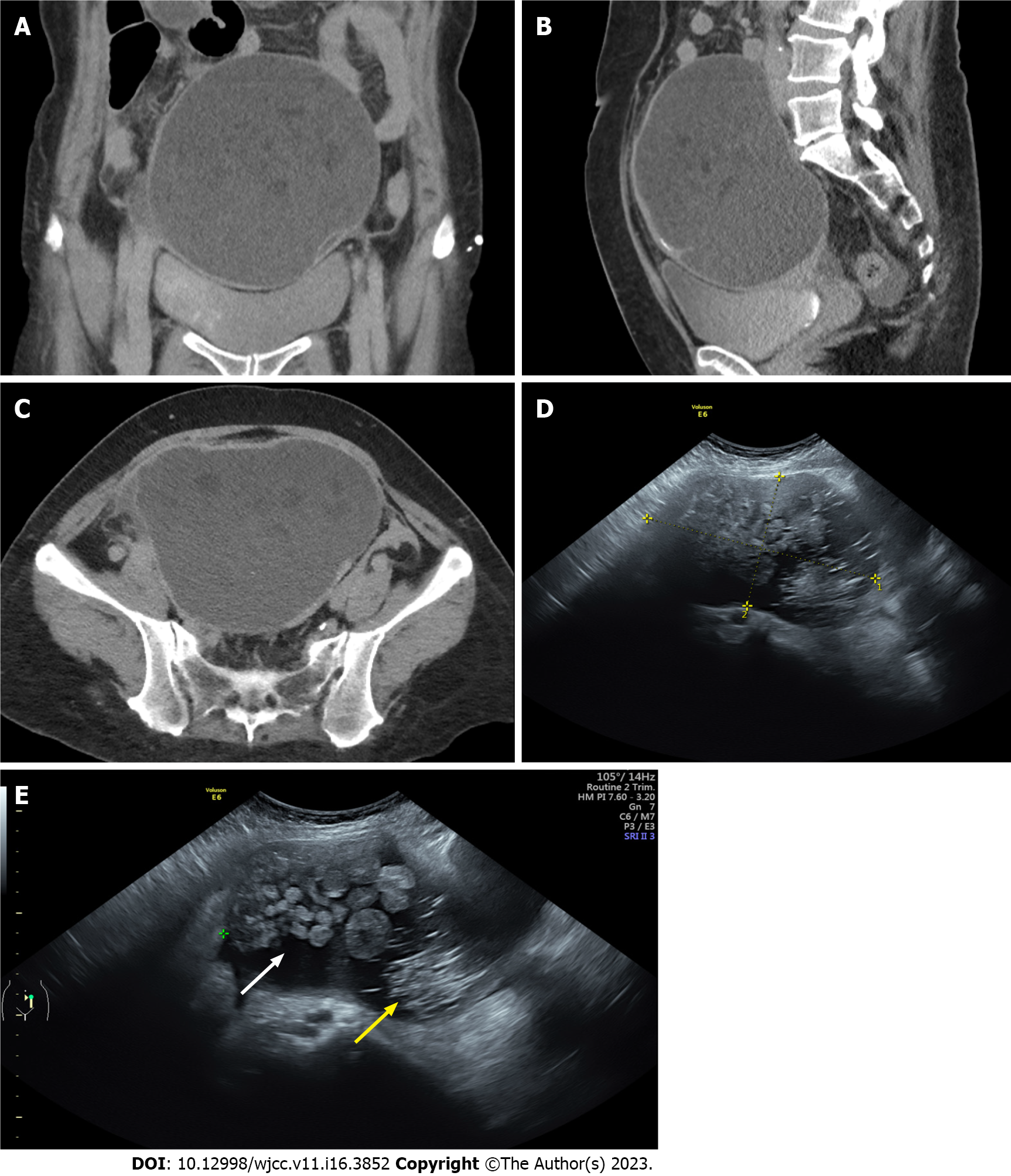
Figure 1 Image studies of the tumor.
A computer tomography scan showing a large tumor with septa in the abdominal cavity. A: Coronary view; B: Sagittal view; C: Axial view; D and E: Pelvic ultrasound showing a tumor with a size of 14.5 cm × 8.6 cm. Hair-strand-like materials (yellow arrow) and papillary-like growth (white arrow) were noted in the tumor.
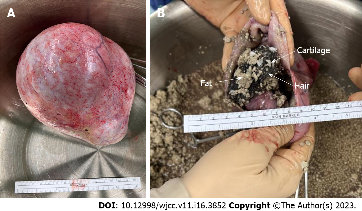
Figure 2 Surgical image of the tumor.
A: Gross picture of the tumor; B: After incising the tumor, the content showed hair-like small ball-like adipose tissues, and cartilage.
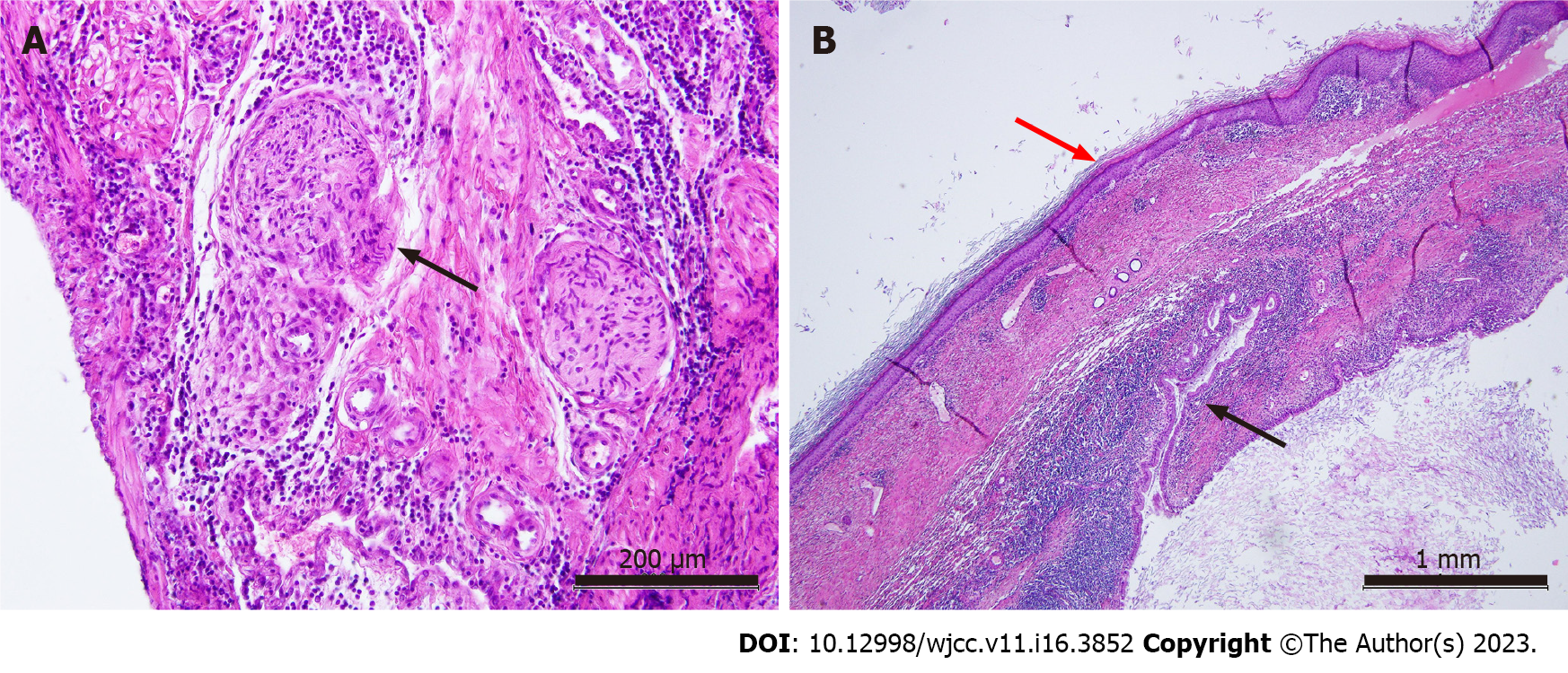
Figure 3 Histopathological study of the tumor (hematoxylin-eosin staining).
A: Nerve fiber (black arrow), scale bar = 200 μm; B: Squamous epithelium (red arrow) and columnar epithelium (intestine, black arrow), scale bar = 1 mm.
- Citation: Lai PH, Ding DC. Ruptured teratoma mimicking a pelvic inflammatory disease and ovarian malignancy: A case report. World J Clin Cases 2023; 11(16): 3852-3857
- URL: https://www.wjgnet.com/2307-8960/full/v11/i16/3852.htm
- DOI: https://dx.doi.org/10.12998/wjcc.v11.i16.3852


