Copyright
©The Author(s) 2023.
World J Clin Cases. Jun 6, 2023; 11(16): 3847-3851
Published online Jun 6, 2023. doi: 10.12998/wjcc.v11.i16.3847
Published online Jun 6, 2023. doi: 10.12998/wjcc.v11.i16.3847
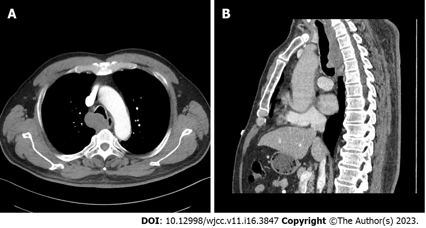
Figure 1 Computed tomography scan of the airway occupation.
A: Chest Computed tomography (CT); B: CT with three-dimensional airway reconstruction.
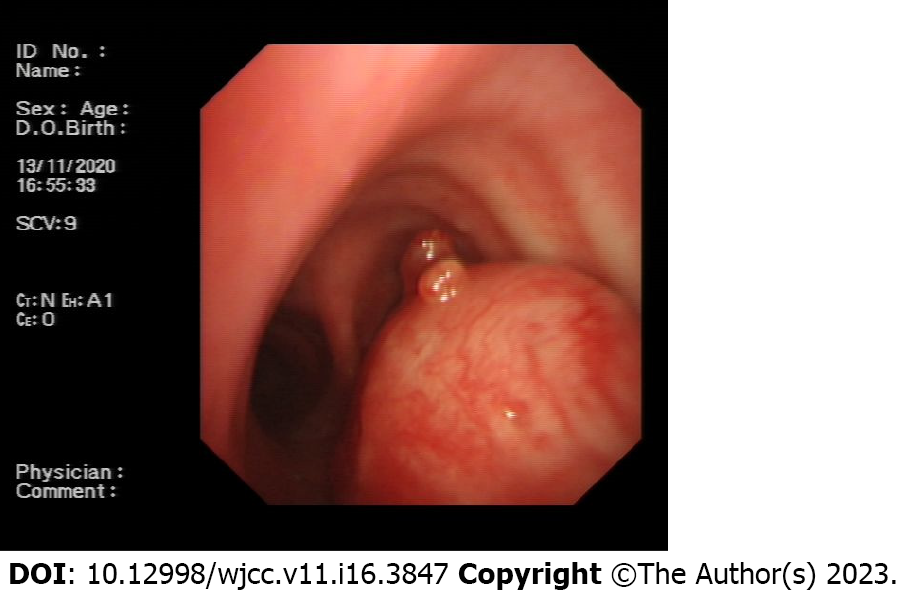
Figure 2 Tracheal bronchoscopy.
The masses originated from the trachea with expansive growth.
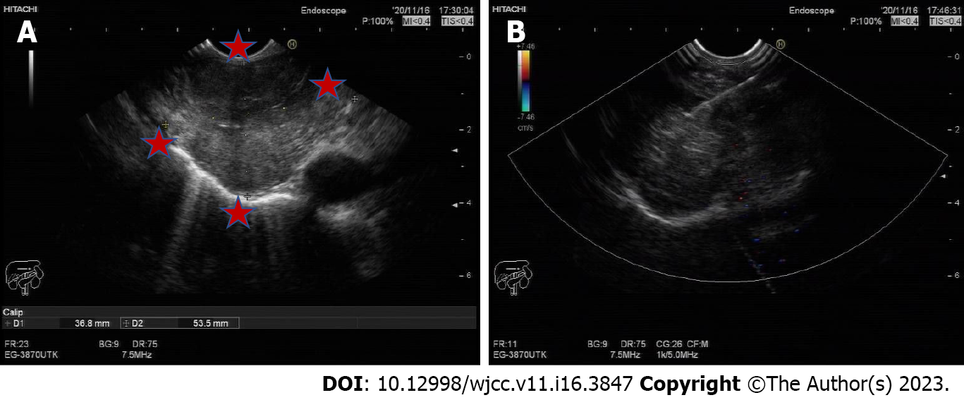
Figure 3 Ultrasound scan.
A and B: A mass with an unclear boundary.
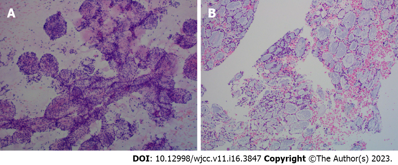
Figure 4 Pathology (100 ×) of the tumour.
A: Cytological pathology; B: Tissue pathology.
- Citation: Pu XX, Xu QW, Liu BY. TACC diagnosed by transoesophageal endoscopic ultrasonography: A case report. World J Clin Cases 2023; 11(16): 3847-3851
- URL: https://www.wjgnet.com/2307-8960/full/v11/i16/3847.htm
- DOI: https://dx.doi.org/10.12998/wjcc.v11.i16.3847


