Copyright
©The Author(s) 2023.
World J Clin Cases. Jun 6, 2023; 11(16): 3822-3829
Published online Jun 6, 2023. doi: 10.12998/wjcc.v11.i16.3822
Published online Jun 6, 2023. doi: 10.12998/wjcc.v11.i16.3822
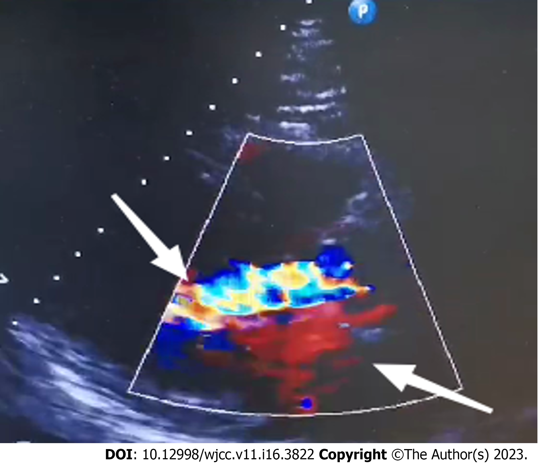
Figure 1
Bidirectional regurgitation of the aortic valve (shown by the arrows).
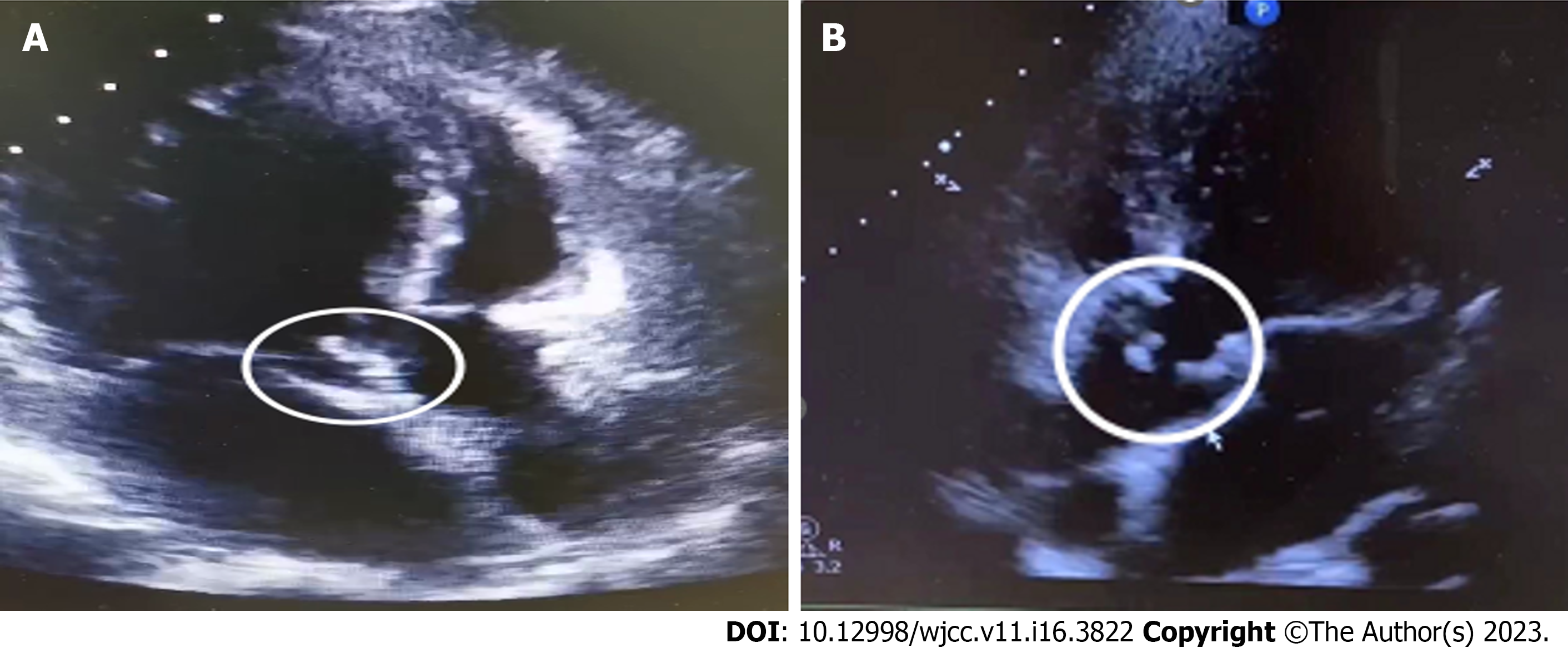
Figure 2 Aortic valve.
A: Aortic valve bulge (shown in circle); B: Aortic valve redundancy detached with perforation (shown in circle).
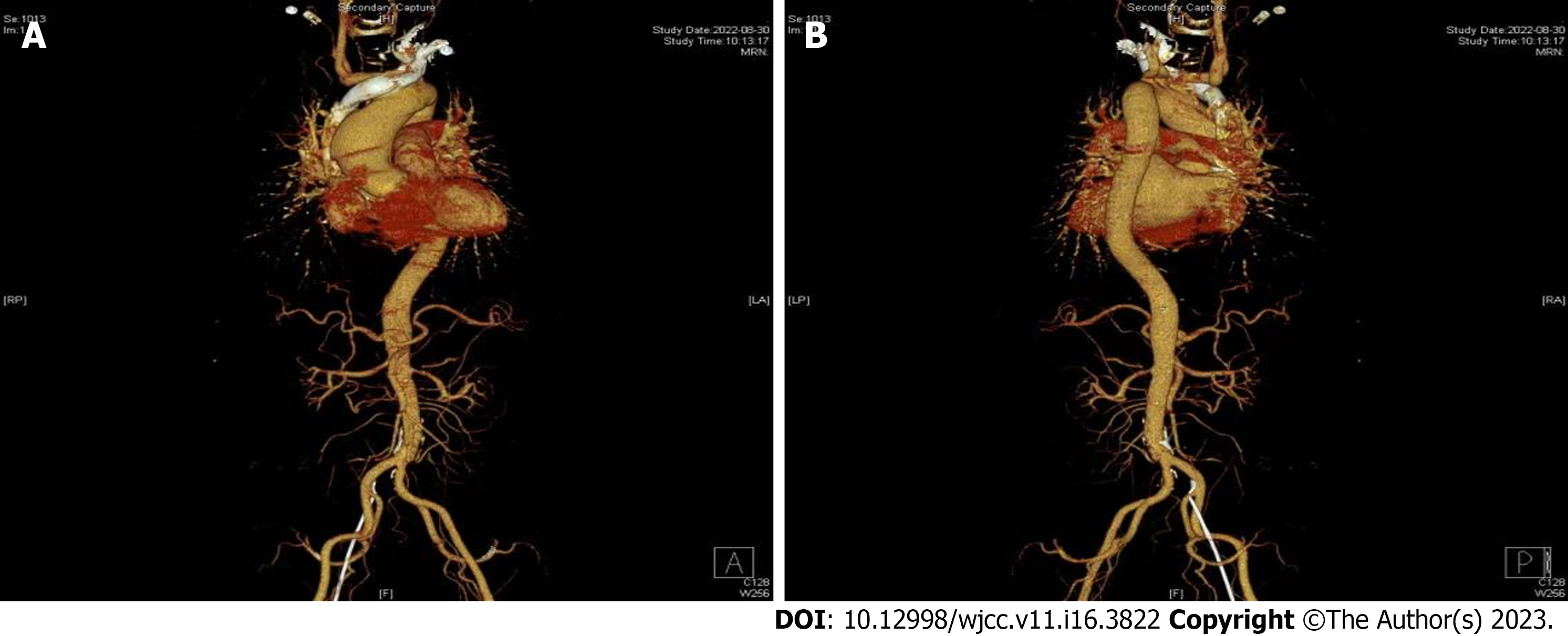
Figure 3 Computed tomography angiography examination results.
A: moderate stenosis of the lumen at the beginning of the abdominal trunk, with possible bacteriophage involvement; B: Visible penetrating ulcers can be seen.
- Citation: Qu YF, Yang J, Wang JY, Wei B, Ye XH, Li YX, Han SL. Valve repair after infective endocarditis secondary to perforation caused by Streptococcus gordonii: A case report. World J Clin Cases 2023; 11(16): 3822-3829
- URL: https://www.wjgnet.com/2307-8960/full/v11/i16/3822.htm
- DOI: https://dx.doi.org/10.12998/wjcc.v11.i16.3822


