Copyright
©The Author(s) 2023.
World J Clin Cases. Jun 6, 2023; 11(16): 3802-3812
Published online Jun 6, 2023. doi: 10.12998/wjcc.v11.i16.3802
Published online Jun 6, 2023. doi: 10.12998/wjcc.v11.i16.3802
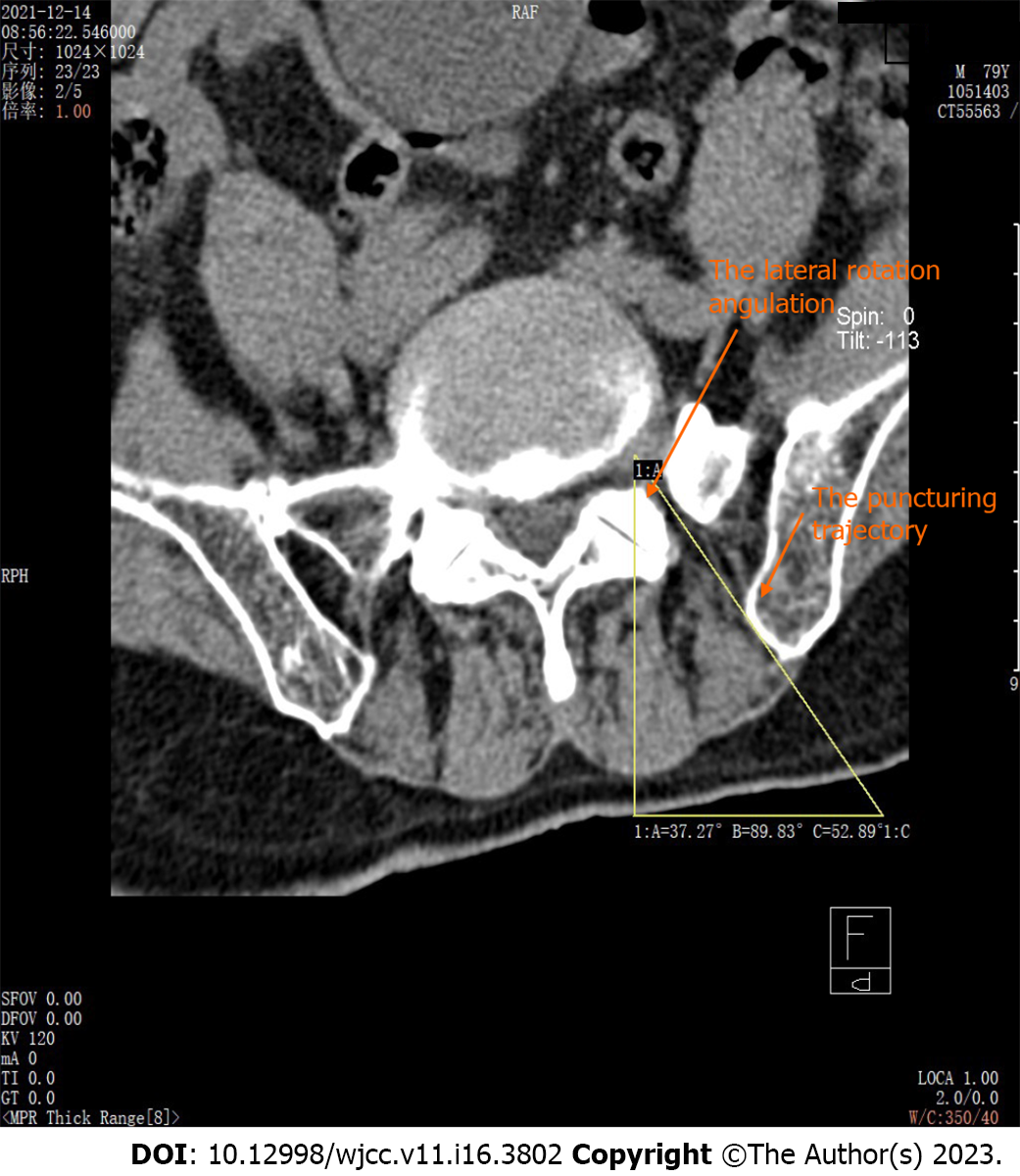
Figure 1 Proper trajectory angle measured in computed tomography scan.
For this far lateral type herniated lumbar disc case, a much smaller lateral tilt angulation of the trajectory was measured in the computed tomography scan beforehand.
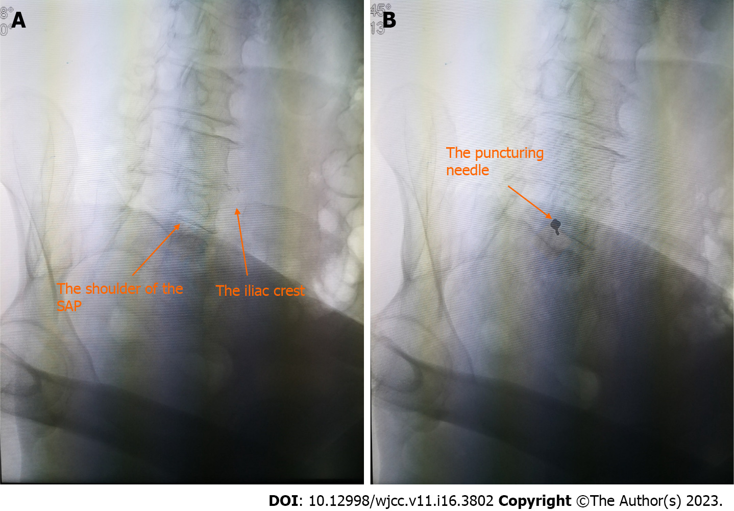
Figure 2 X-ray films of the coaxial radiography-guided puncture technique.
A: For the coaxial radiography-guided puncture technique, the target should be identified first, which is usually the shoulder of the superior articular process; B: The needle was maintained coaxial to the X-ray beam and parallel to the X-ray axis. In this instance, the needle hub projected directly over the tip and aligned with the target, and the needle appeared as a dot instead of a line. SAP: Superior articular process.
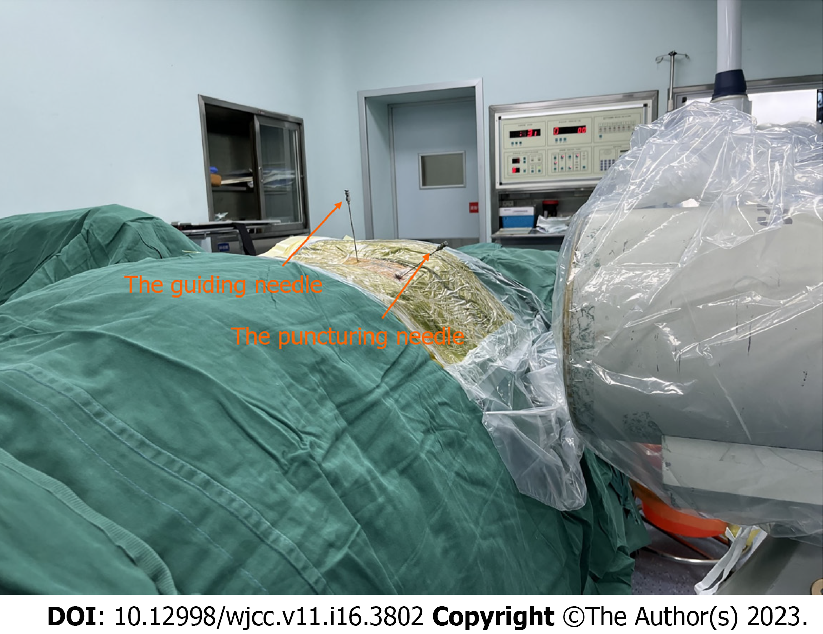
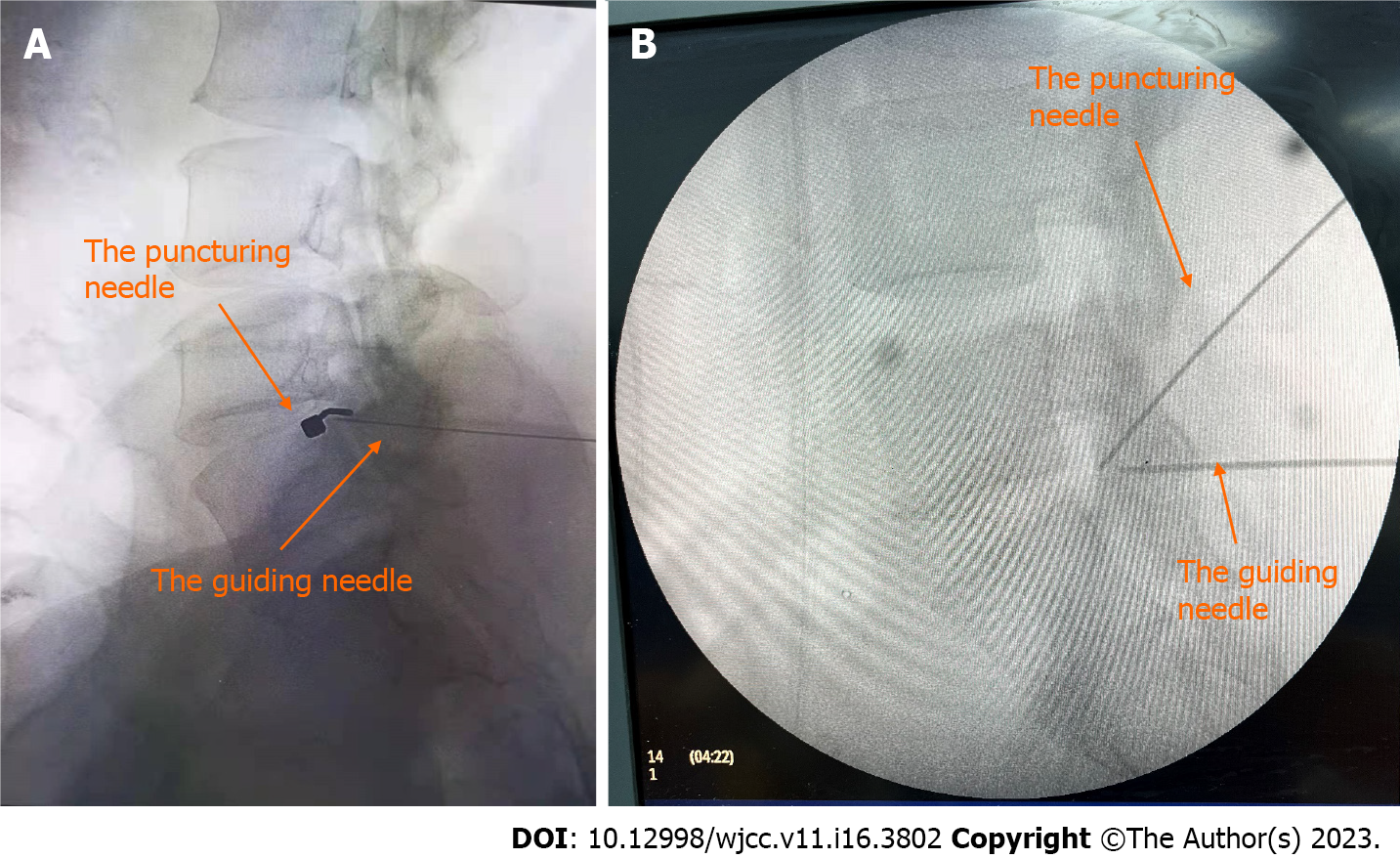
Figure 3 Picture of the coaxial radiography-guided puncture technique with an additional guiding needle.
If the superior articular process could not be visualized clearly, a guiding needle was used in addition to the puncturing needle. This usually occurred when the lateral oblique angle of the C-arm was very large.
Figure 4 X-ray films of a successful puncturing of a guiding needle.
The anteroposterior and lateral views confirmed the needle tip placement near the ventral margin of the superior articular process.
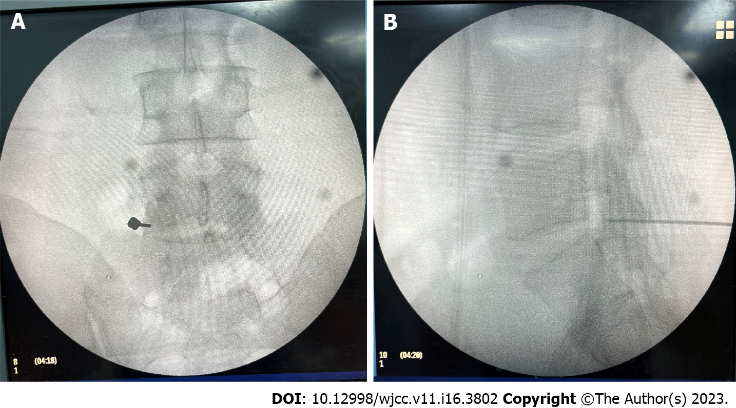
Figure 5 X-ray films of a successful puncturing with an additional guiding needle.
A: The C-arm was set at the coaxial position and puncture was performed under coaxial radiography guidance. In this position, the puncturing needle was shown as a dot rather than a line; B: The lateral view shows that the puncturing needle has reached the tip of the guiding needle, indicating a successful puncture.
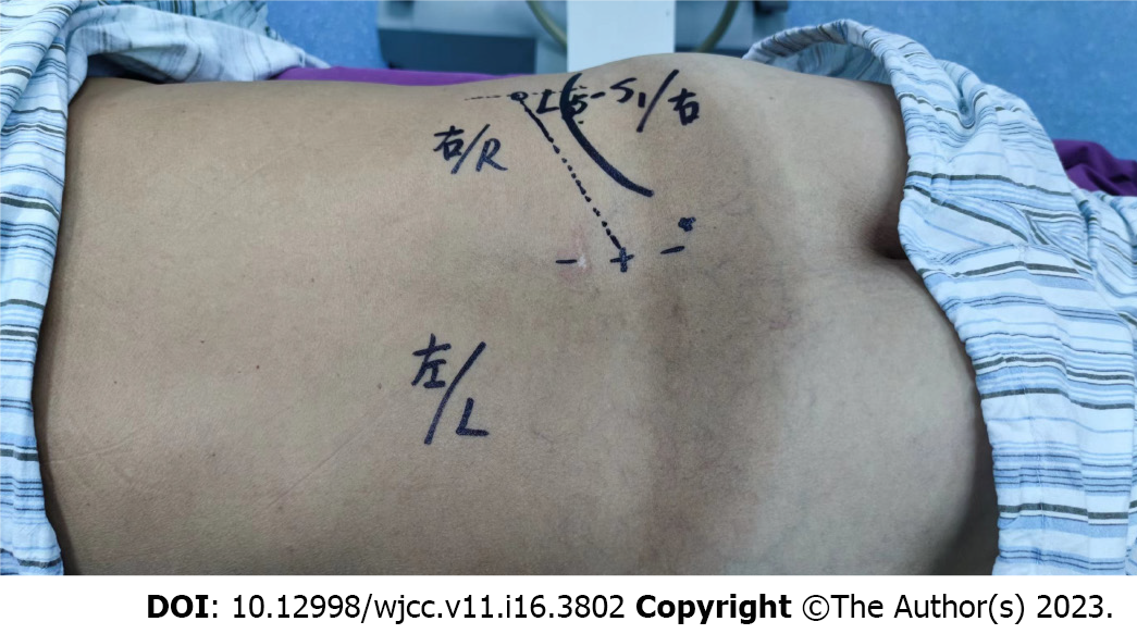
Figure 6 Picture of puncturing direction in the radiography-guided puncture technique marked in the skin.
A lateral longitudinal line parallel to the center longitudinal line was drawn, about 10-14 cm from the center line, and the intersection of the lateral longitudinal line and the oblique line passing through the tip of the superior articular process was chosen as the puncture point.
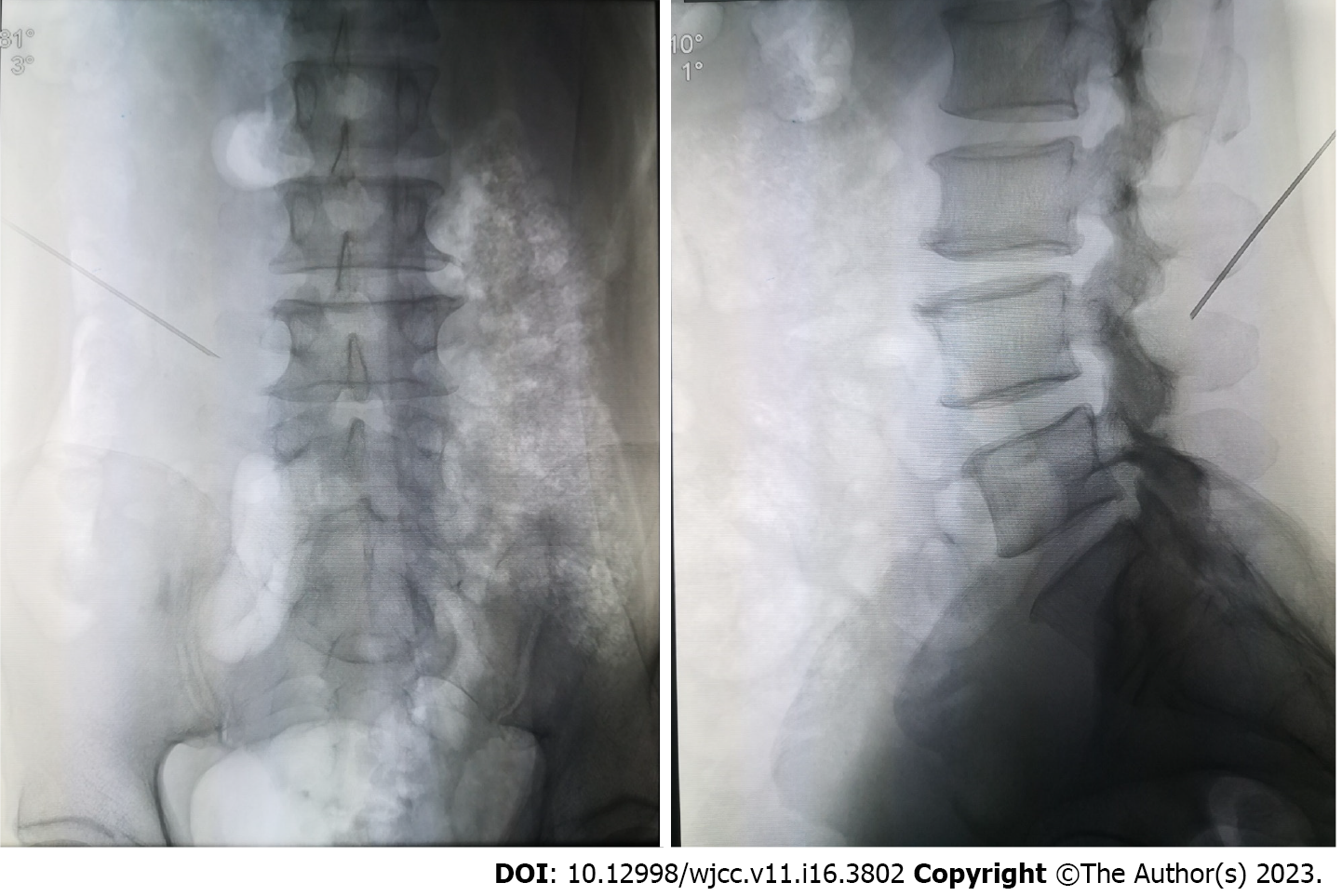
Figure 7 X-ray films of the radiography-guided puncture technique.
The needle should be oriented to the shoulder of the superior articular process under both the anteroposterior and lateral views.
- Citation: Chen LP, Wen BS, Xu H, Lu Z, Yan LJ, Deng H, Fu HB, Yuan HJ, Hu PP. Coaxial radiography guided puncture technique for percutaneous transforaminal endoscopic lumbar discectomy: A randomized control trial. World J Clin Cases 2023; 11(16): 3802-3812
- URL: https://www.wjgnet.com/2307-8960/full/v11/i16/3802.htm
- DOI: https://dx.doi.org/10.12998/wjcc.v11.i16.3802


