Copyright
©The Author(s) 2023.
World J Clin Cases. Jun 6, 2023; 11(16): 3680-3693
Published online Jun 6, 2023. doi: 10.12998/wjcc.v11.i16.3680
Published online Jun 6, 2023. doi: 10.12998/wjcc.v11.i16.3680
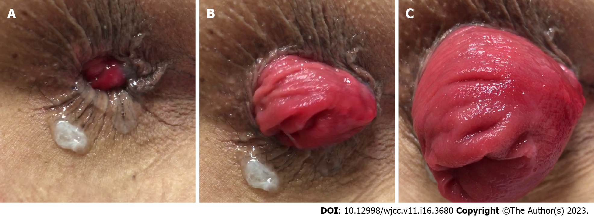
Figure 1 Progression of external prolapse out of the anal canal.
A-C: Prolapse becomes more evident with straining.
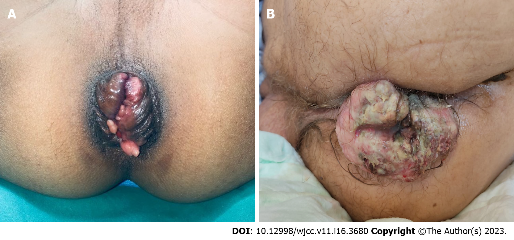
Figure 2 Differential diagnoses of rectal prolapse.
A: Prolapsing hemorrhoids; B: Anal canal mass.
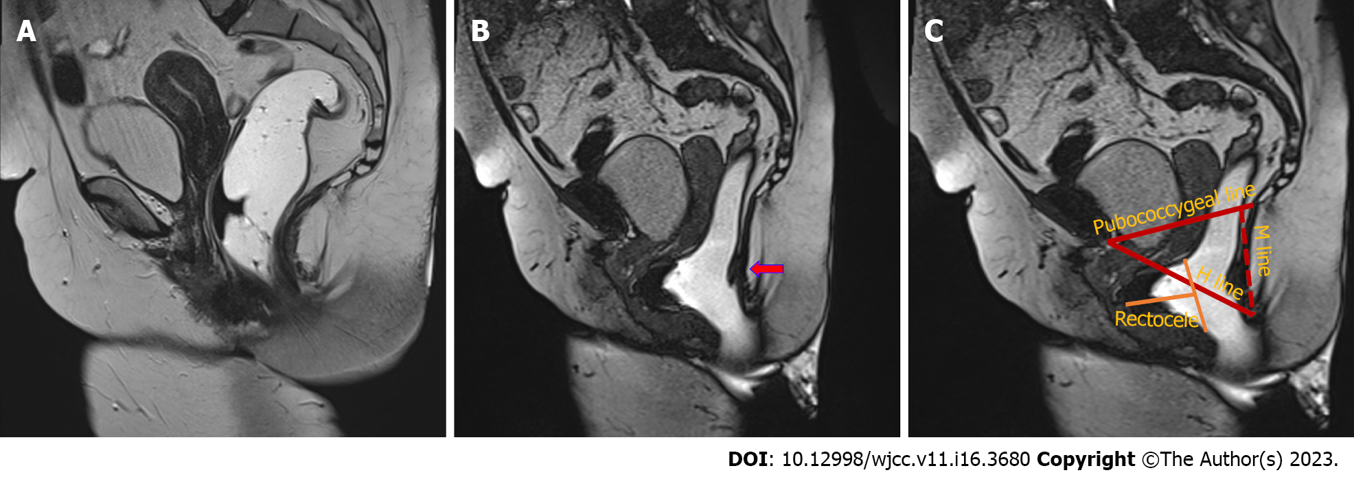
Figure 3 Dynamic magnetic resonance defecography images.
A: Magnetic resonance imaging during rest; B: The red arrow indicates slight rectal intussusception and advanced pelvic prolapse present during straining; C: Pubococcygeal, M and H lines. The yellow line reveals severe rectocele accompanying pelvic prolapse.
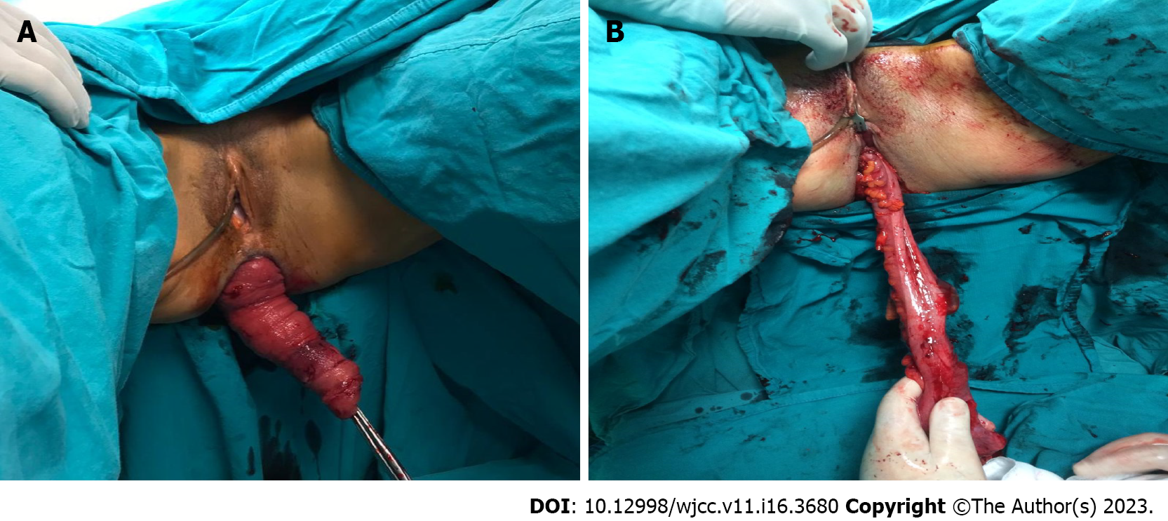
Figure 4 A patient with external rectal prolapse who underwent an alternative procedure.
A: External rectal prolapse; B: Altemeier procedure.
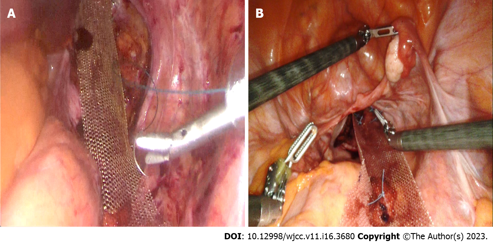
Figure 5 Laparoscopic and robotic ventral mesh rectopexy.
A: Laparoscopic mesh placement before peritoneal closure; B: Robotic mesh placement before peritoneal closure.
- Citation: Oruc M, Erol T. Current diagnostic tools and treatment modalities for rectal prolapse. World J Clin Cases 2023; 11(16): 3680-3693
- URL: https://www.wjgnet.com/2307-8960/full/v11/i16/3680.htm
- DOI: https://dx.doi.org/10.12998/wjcc.v11.i16.3680


