Copyright
©The Author(s) 2023.
World J Clin Cases. May 26, 2023; 11(15): 3643-3650
Published online May 26, 2023. doi: 10.12998/wjcc.v11.i15.3643
Published online May 26, 2023. doi: 10.12998/wjcc.v11.i15.3643
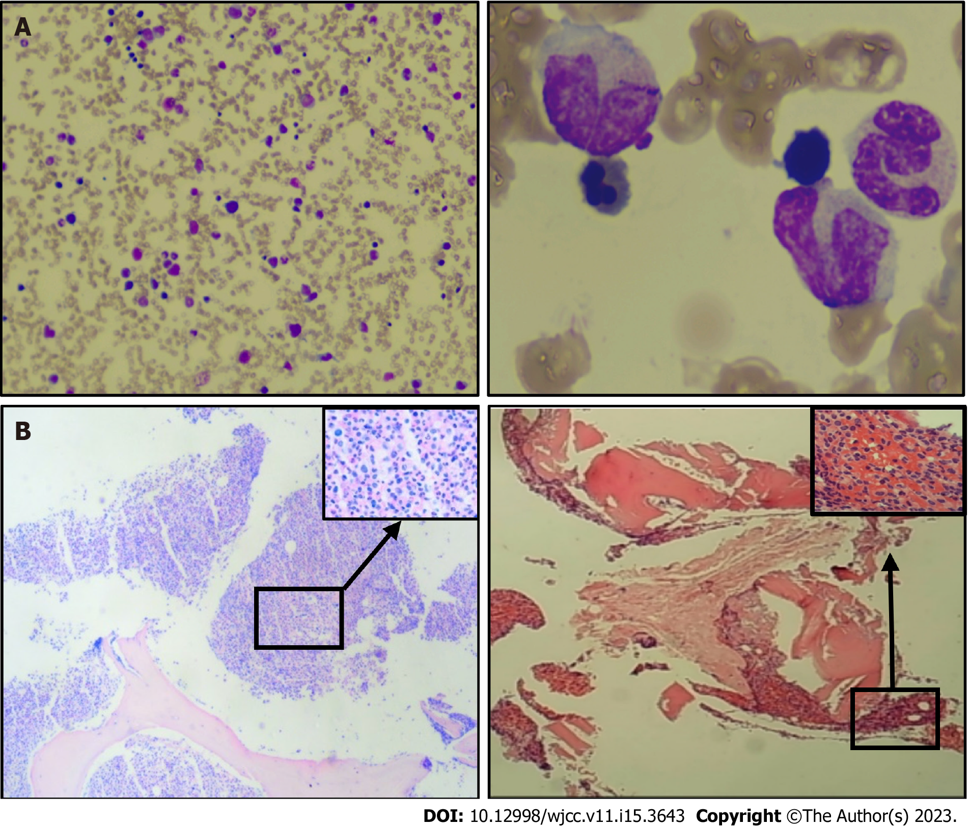
Figure 1 Morphological and biopsy results of chronic myelomonocytic leukemia bone marrow analysis.
A: Pelger deformed granules (100× magnification), exhibiting abnormal proliferation of monocytes and occasional naïve monocytes (with well-differentiated monocytes accounting for 45% of total monocytes), inhibited erythroid hyperplasia, three megakaryocytes, and rare platelet; B: Increased naïve-stage cells, with an increased granulocyte-to-nucleated red blood cell ratio. The granular lineage predominantly comprised intermediate (lower-stage) and visible monocytes, based on reticulocyte staining (MF-0 grade; ×4 and ×40 magnification).
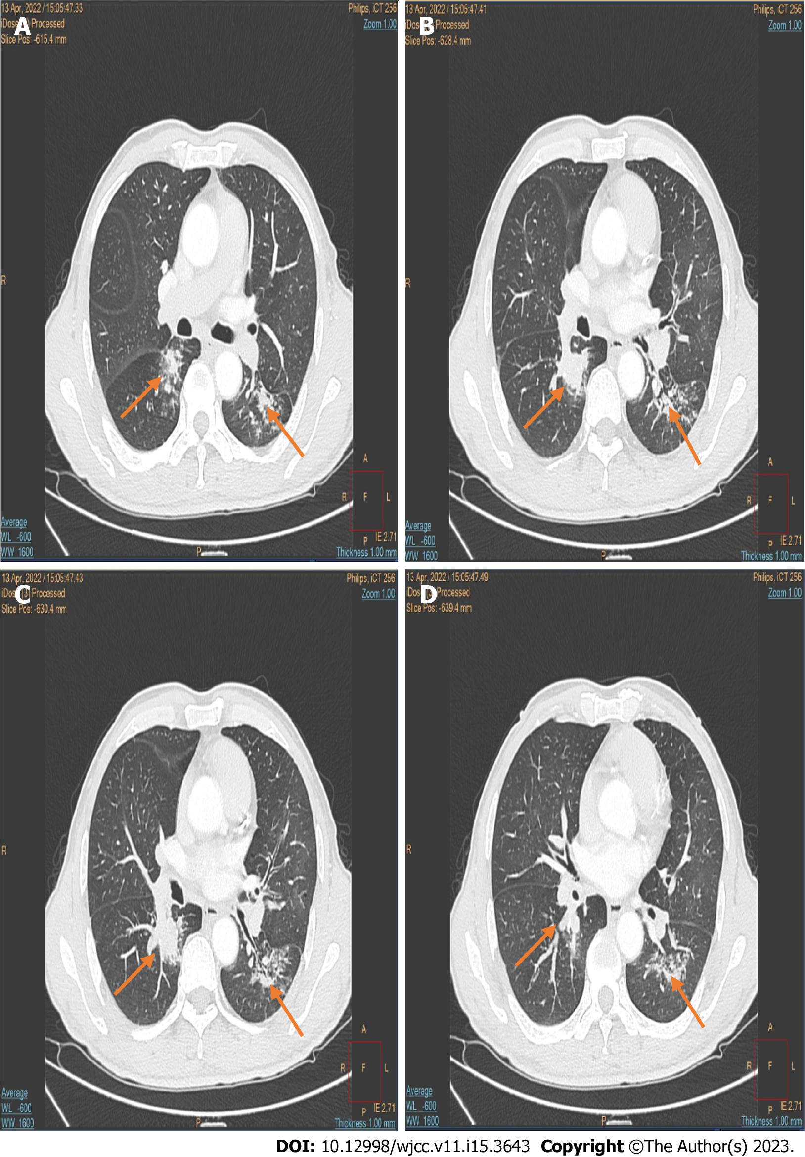
Figure 2 Enhanced computed tomography results for non-small cell lung cancer.
A: Squamous cell carcinoma, showing soft tissue density in the dorsal segment of the right lower lobe of the lung and inflammatory lesions in both lungs; B: Next slice chest enhanced computed tomography (CT) image; C: Next slice chest enhanced CT image; D: Next slice chest enhanced CT image.
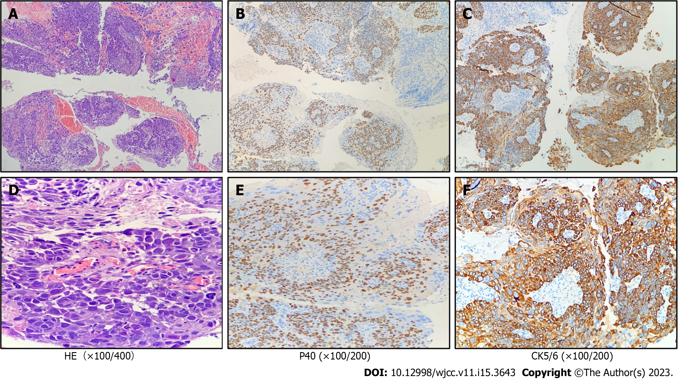
Figure 3 Immunohistochemistry results and pathological manifestations of non-small cell lung cancer squamous cell carcinoma.
A: HE staining was used to observe the pathological manifestation (×100 magnification); B: Nuclear positive (P40, ×100 magnification); C: Cytoplasmic positive (CK5/6, ×100 magnification); D: Tumor cells with large and deeply stained nuclei, significant nucleoplasmic heterogeneity, keratinization in some areas, rich blood vessels, tumor cells growing around blood vessels, and polarity disappearing in some areas, showing solid nests (HE, ×400 magnification); E: Nuclear positive (P40, ×200 magnification); F: Cytoplasmic positive (CK5/6, ×200 magnification).
- Citation: Deng LJ, Dong Y, Li MM, Sun CG. Co-existing squamous cell carcinoma and chronic myelomonocytic leukemia with ASXL1 and EZH2 gene mutations: A case report. World J Clin Cases 2023; 11(15): 3643-3650
- URL: https://www.wjgnet.com/2307-8960/full/v11/i15/3643.htm
- DOI: https://dx.doi.org/10.12998/wjcc.v11.i15.3643


