Copyright
©The Author(s) 2023.
World J Clin Cases. May 26, 2023; 11(15): 3631-3636
Published online May 26, 2023. doi: 10.12998/wjcc.v11.i15.3631
Published online May 26, 2023. doi: 10.12998/wjcc.v11.i15.3631
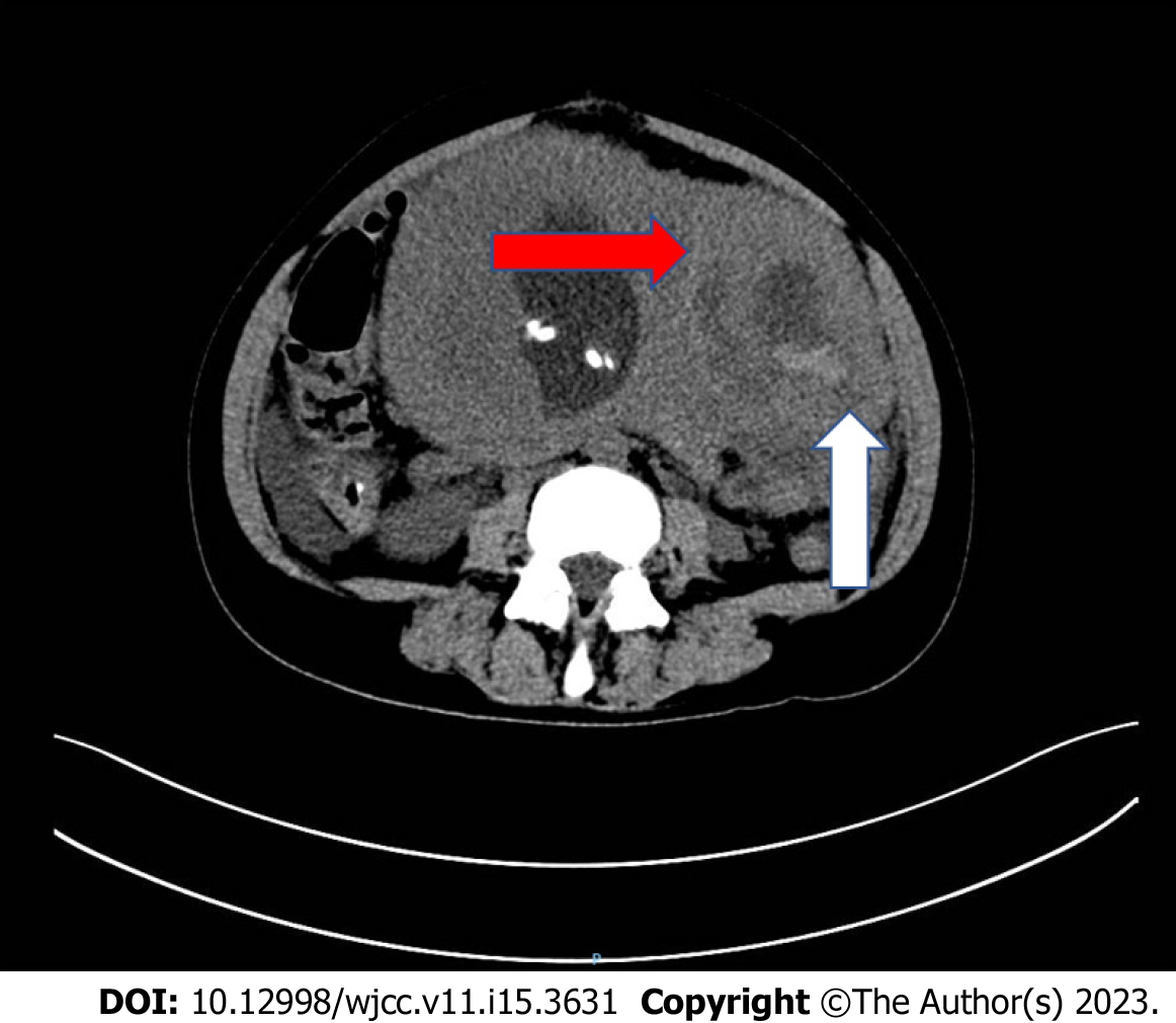
Figure 1 Computed tomography of the abdomen showing a 10.
9 cm × 7.1 cm × 8.3 cm degenerated fibroid (red arrow) and myoma laceration (white arrow).
Figure 2 Laparoscopic exploration.
A: Laparoscopic exploration revealed rupture of hysteromyoma, and the rupture of hysteromyoma was covered by the greater omentum; B: Laparoscopic exploration revealed hematocele of hepatic region.
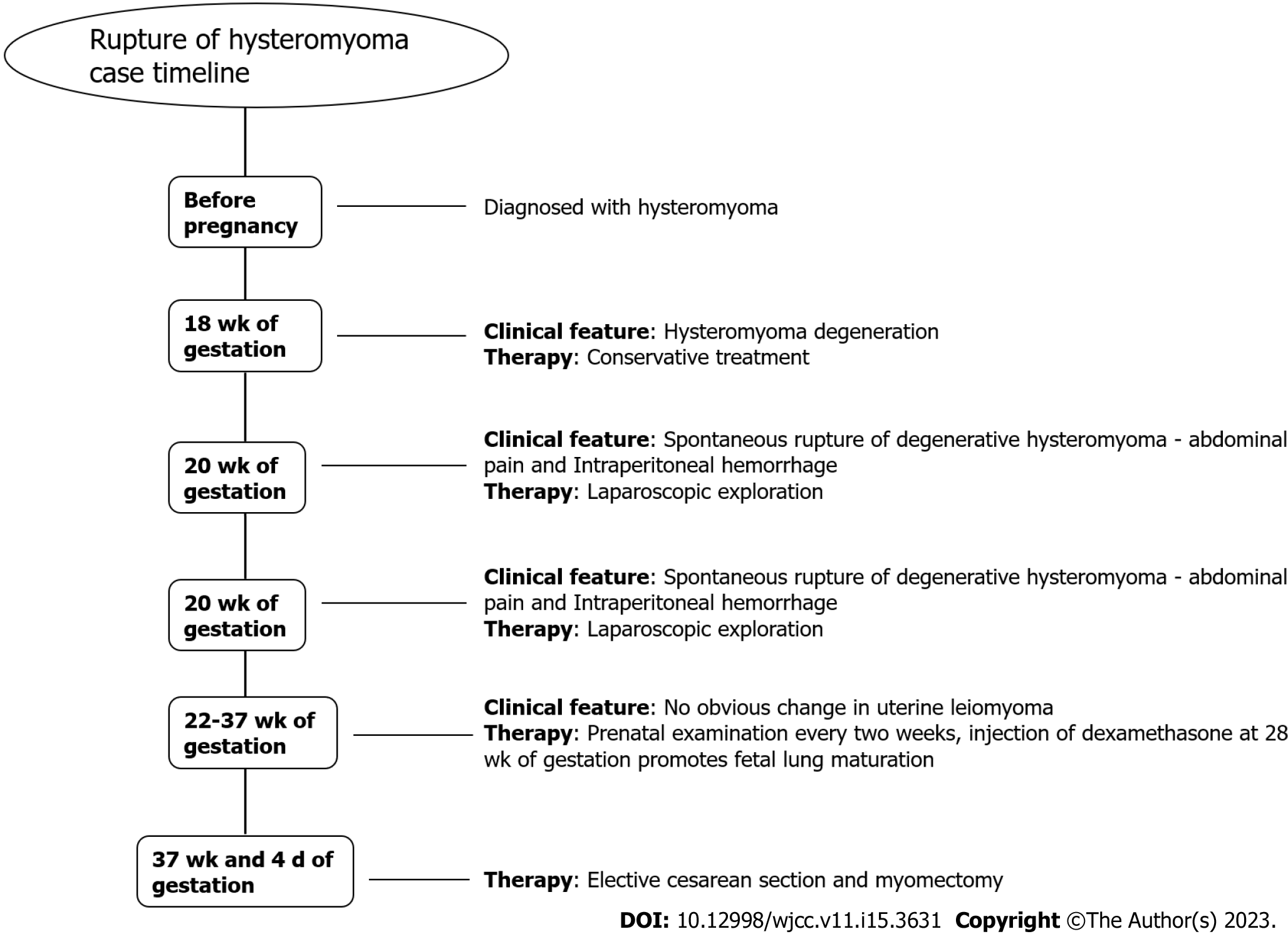
Figure 3 Timeline of diagnosis and treatment of the patient.
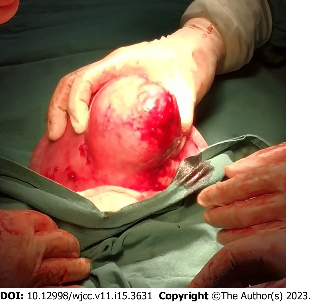
Figure 4 Rupture of the upper part of 10 cm exophytic red degenerated fibroid capsule during cesarean section.
- Citation: Xu Y, Shen X, Pan XY, Gao S. Acute abdomen caused by spontaneous rupture of degenerative hysteromyoma during pregnancy: A case report. World J Clin Cases 2023; 11(15): 3631-3636
- URL: https://www.wjgnet.com/2307-8960/full/v11/i15/3631.htm
- DOI: https://dx.doi.org/10.12998/wjcc.v11.i15.3631


