Copyright
©The Author(s) 2023.
World J Clin Cases. May 26, 2023; 11(15): 3599-3611
Published online May 26, 2023. doi: 10.12998/wjcc.v11.i15.3599
Published online May 26, 2023. doi: 10.12998/wjcc.v11.i15.3599
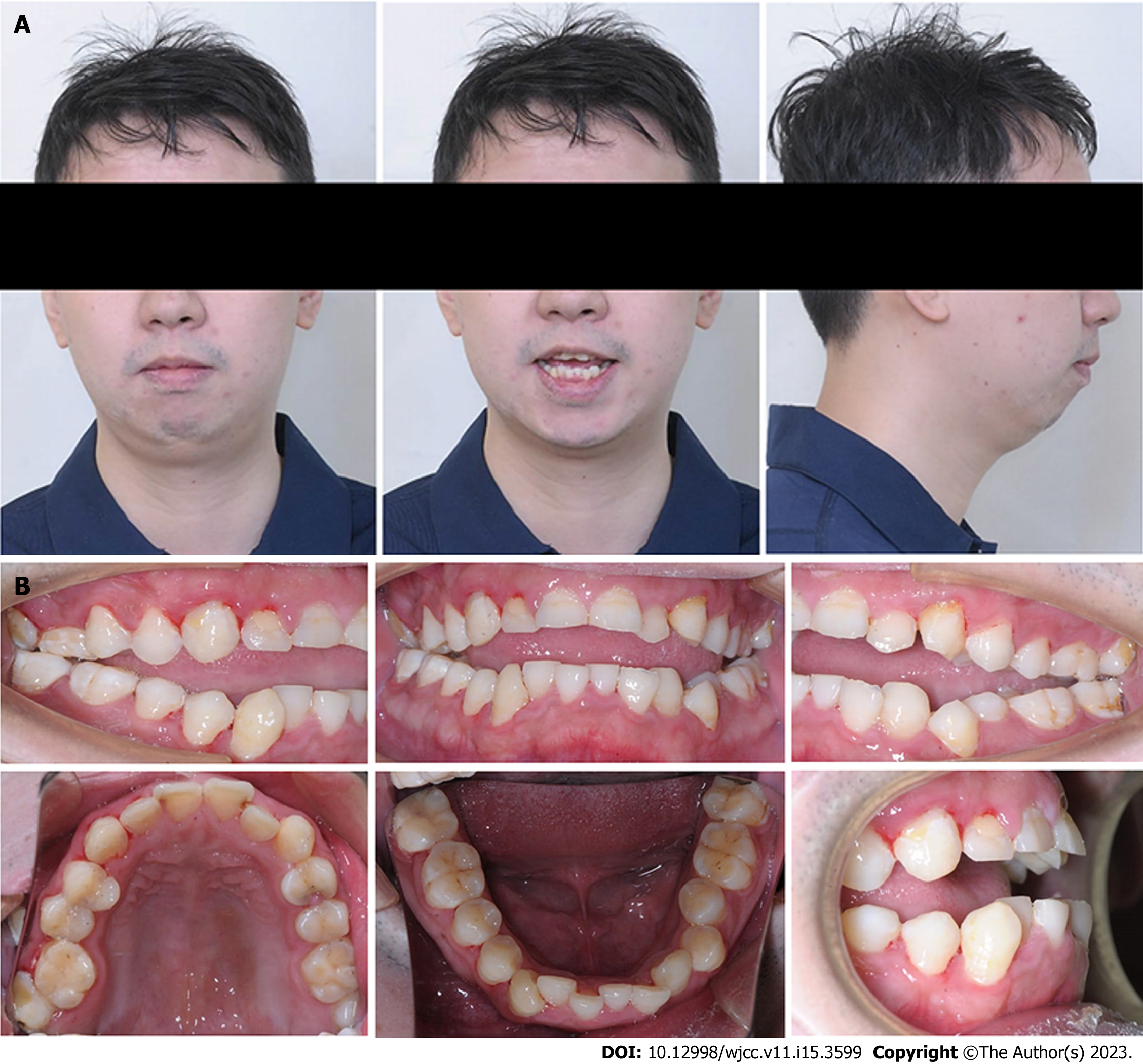
Figure 1 Pretreatment facial and intraoral photographs.
A: Facial photographs; B: Intraoral photographs.
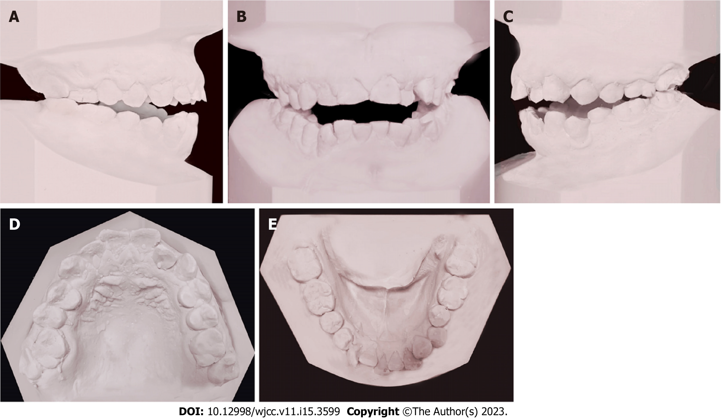
Figure 2 Pretreatment dental models.
A: Right lateral view; B: Frontal view; C: Left lateral view; D: Upper occlusal view; E: Lower occlusal view.
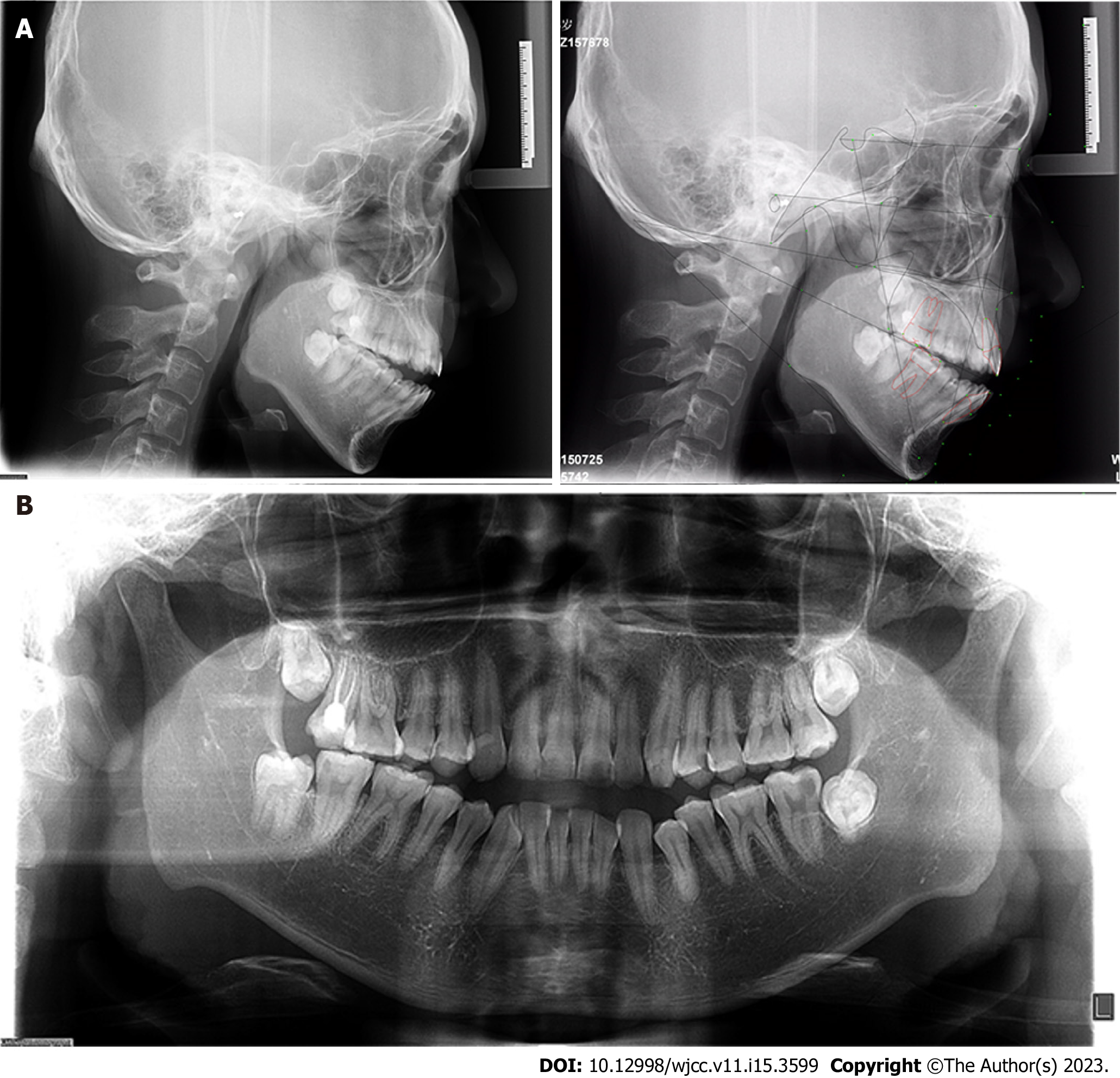
Figure 3 Pretreatment lateral cephalometric and panoramic radiographs.
A: Lateral cephalometric radiographs and tracing; B: Panoramic radiograph.
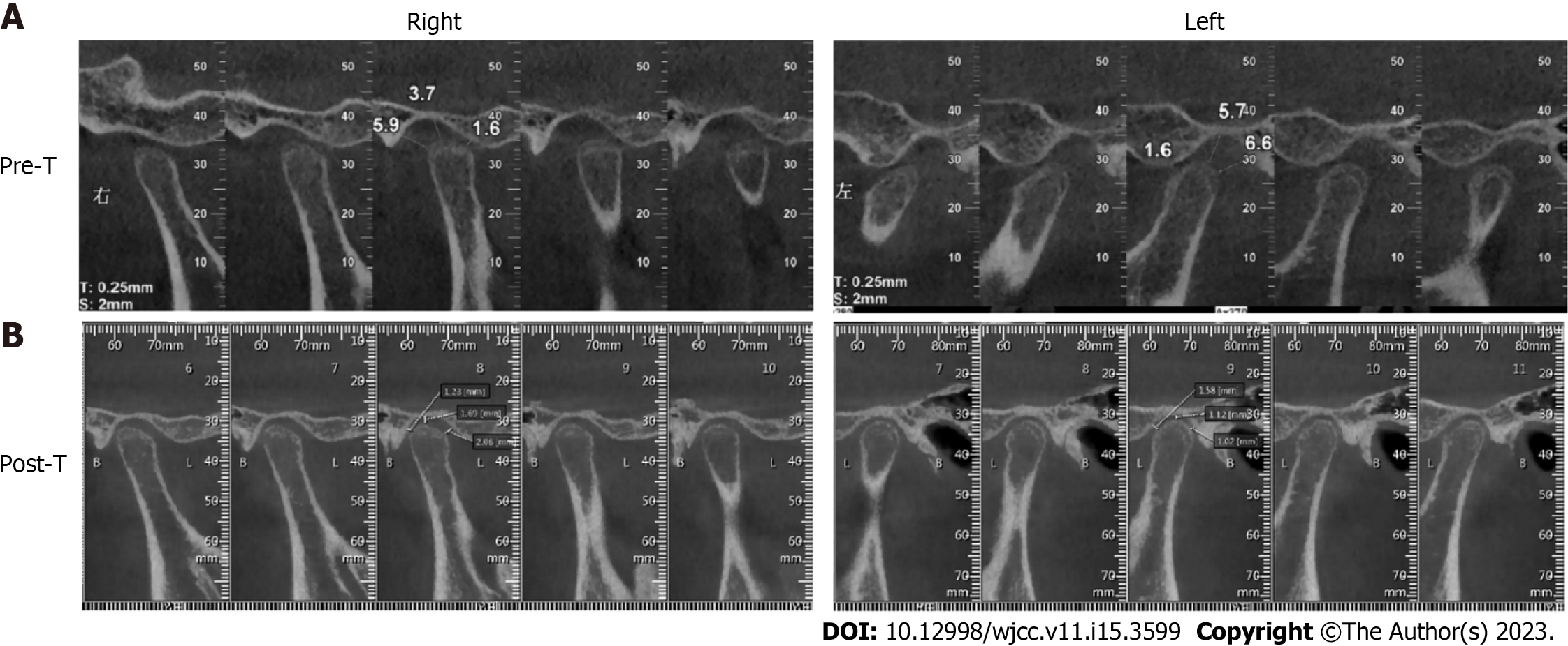
Figure 4 Cone-beam computed tomography images of the temporomandibular joint.
A: Pretreatment; B: Posttreatment.
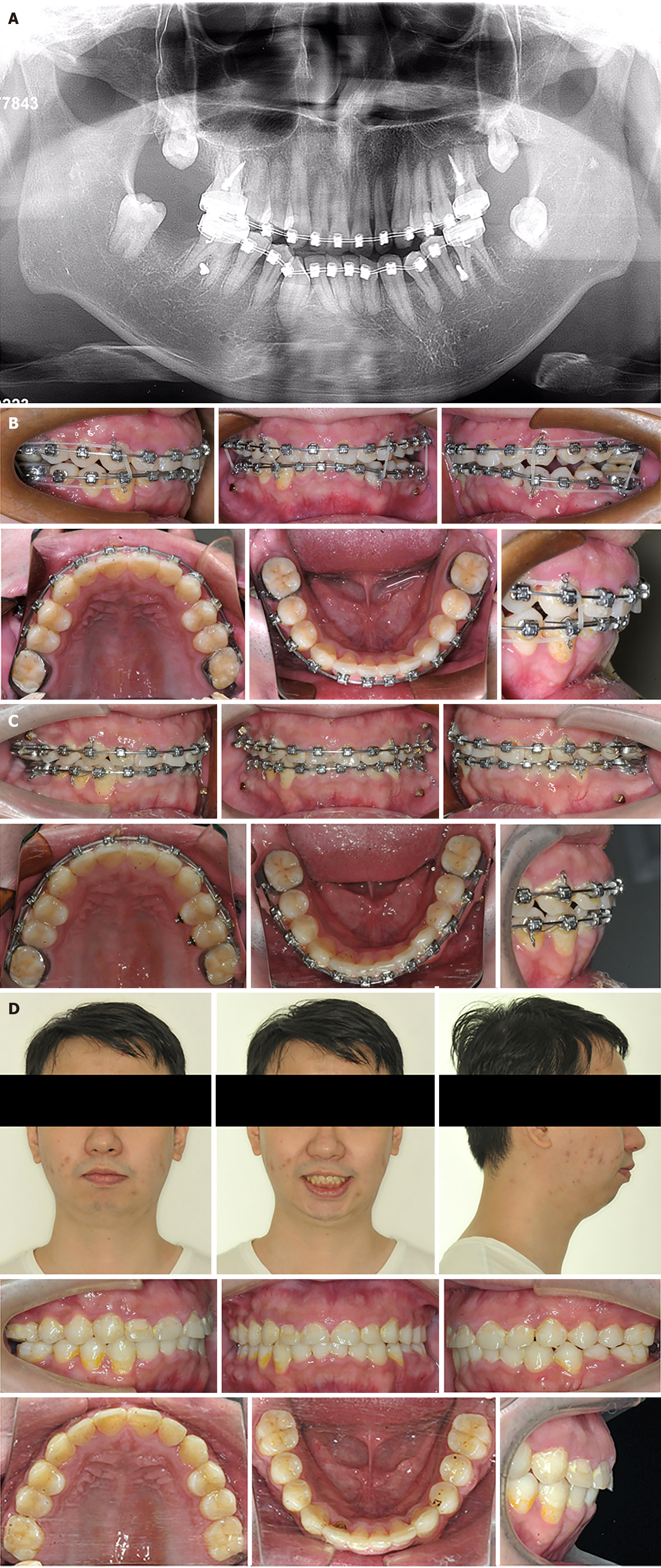
Figure 5 Treatment progress.
A: 2 mo. The four second molars were extracted and 4 mini-implants were implanted to intrude the second premolars and the first molars;B: 8 mo. The anterior open bite was corrected but bilateral posterior open bite occurred; C: 17 mo. After fine adjustment, Class I molar relationship and normal overjet and overbite were obtained; D: Posttreatment facial and intraoral photographs.
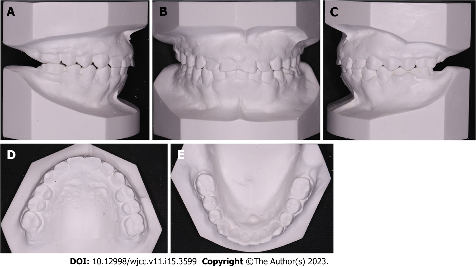
Figure 6 Posttreatment dental models.
A: Right lateral view; B: Frontal view; C: Left lateral view; D: Upper occlusal view; E: Lower occlusal view.
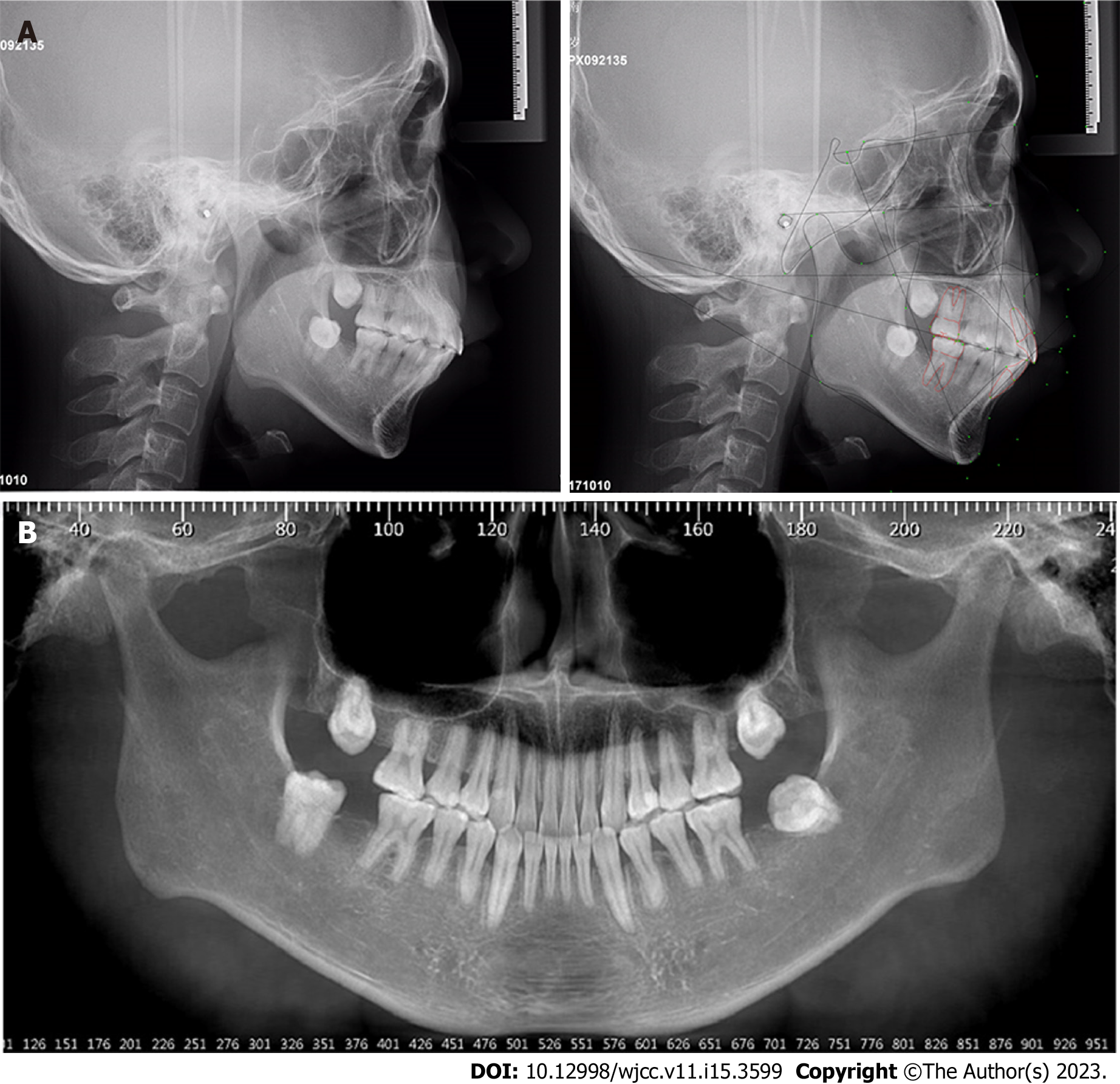
Figure 7 Posttreatment lateral cephalometric and cone-beam computed tomography reconstructed panoramic radiographs.
A: Lateral cephalometric radiographs and tracing; B: Cone-beam computed tomography reconstructed panoramic radiographs.
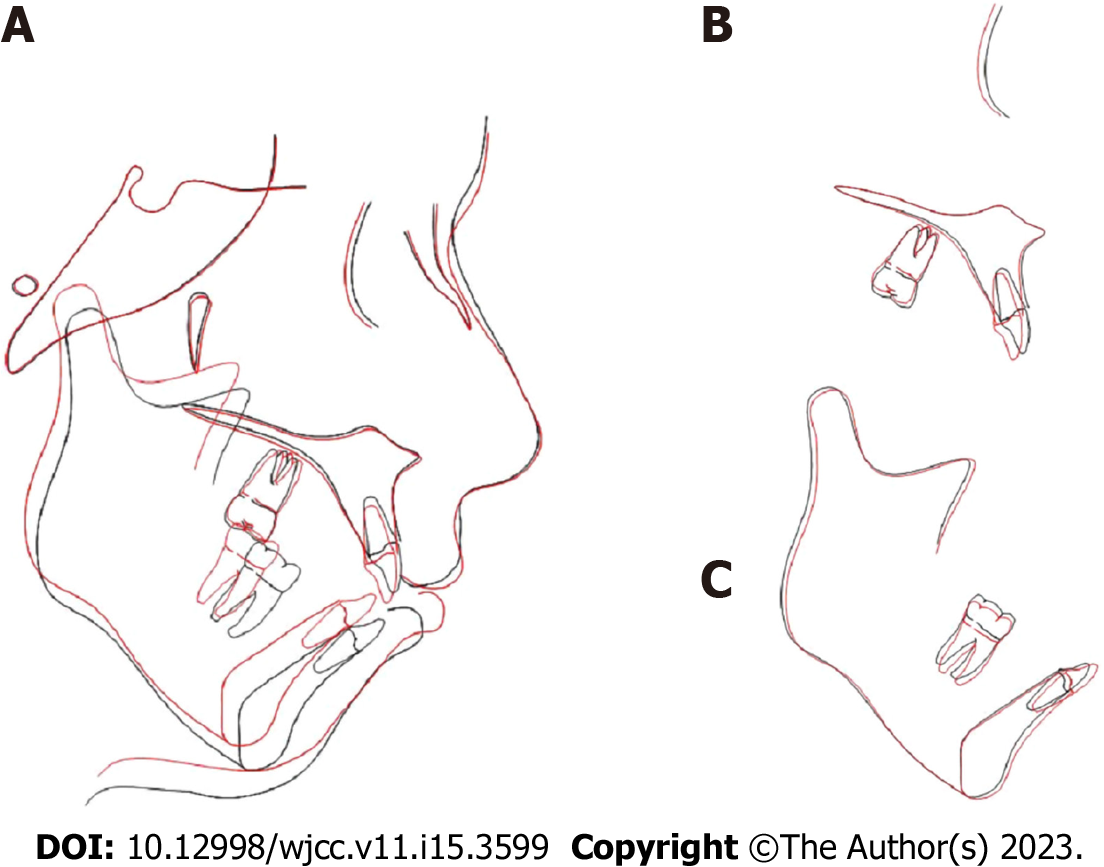
Figure 8 Cephalometric superimpositions.
A: Overall superimposition; B: Maxillary superimposition; C: Mandibular superimposition. Black: Pretreatment; Red: Posttreatment.
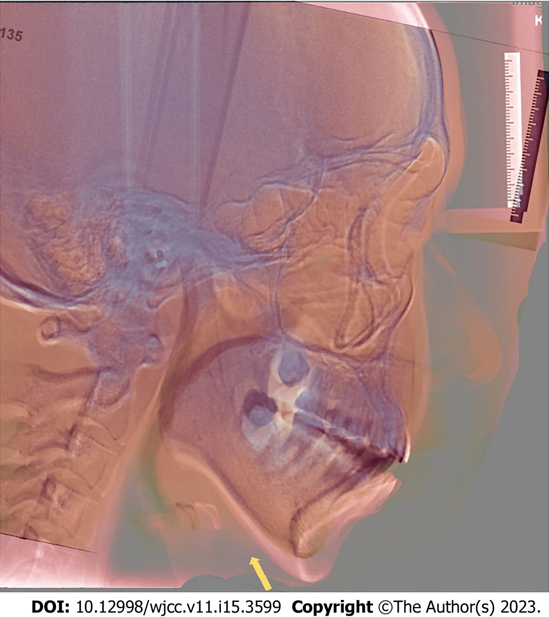
Figure 9 Lateral cephalometric radiographs superimposition.
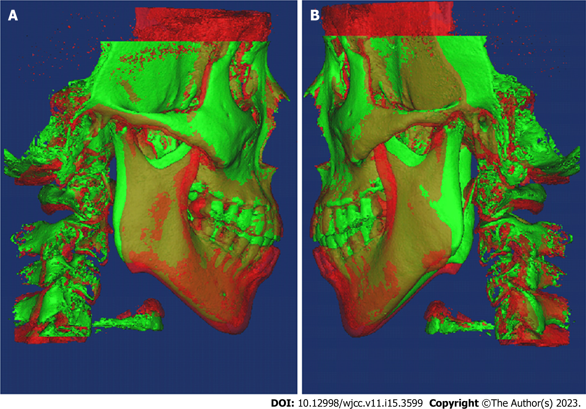
Figure 10 Cone-beam computed tomography 3D superimposition.
A: Right side; B: Left side. Red: Pretreatment; Green: Posttreatment.
- Citation: Huang ZW, Yang R, Gong C, Zhang CX, Wen J, Li H. Treatment of severe open bite and mandibular condyle anterior displacement by mini-screws and four second molars extraction: A case report. World J Clin Cases 2023; 11(15): 3599-3611
- URL: https://www.wjgnet.com/2307-8960/full/v11/i15/3599.htm
- DOI: https://dx.doi.org/10.12998/wjcc.v11.i15.3599


