Copyright
©The Author(s) 2023.
World J Clin Cases. May 26, 2023; 11(15): 3560-3570
Published online May 26, 2023. doi: 10.12998/wjcc.v11.i15.3560
Published online May 26, 2023. doi: 10.12998/wjcc.v11.i15.3560
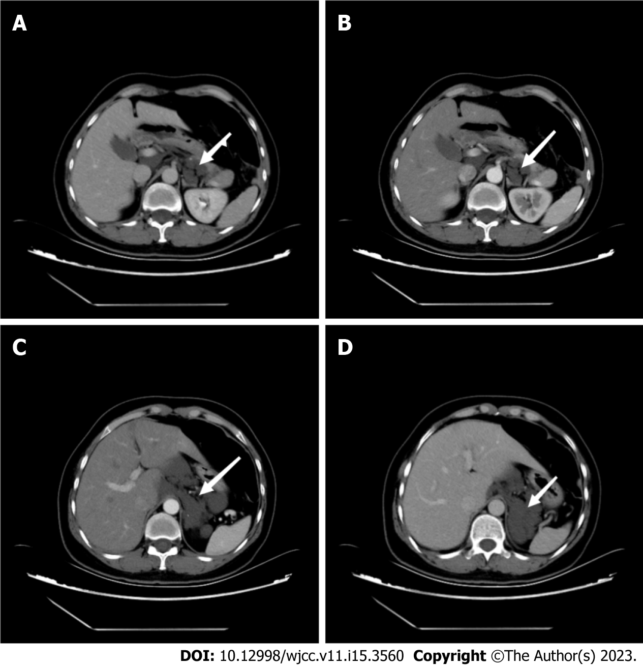
Figure 1 Computed tomography images.
A: Computed tomography showed multiple retroperitoneal cystoid hypodensity shadows (arrow); B-D: No significant enhancement was observed in the arterial phase, balance phase, and venous phase (arrows).
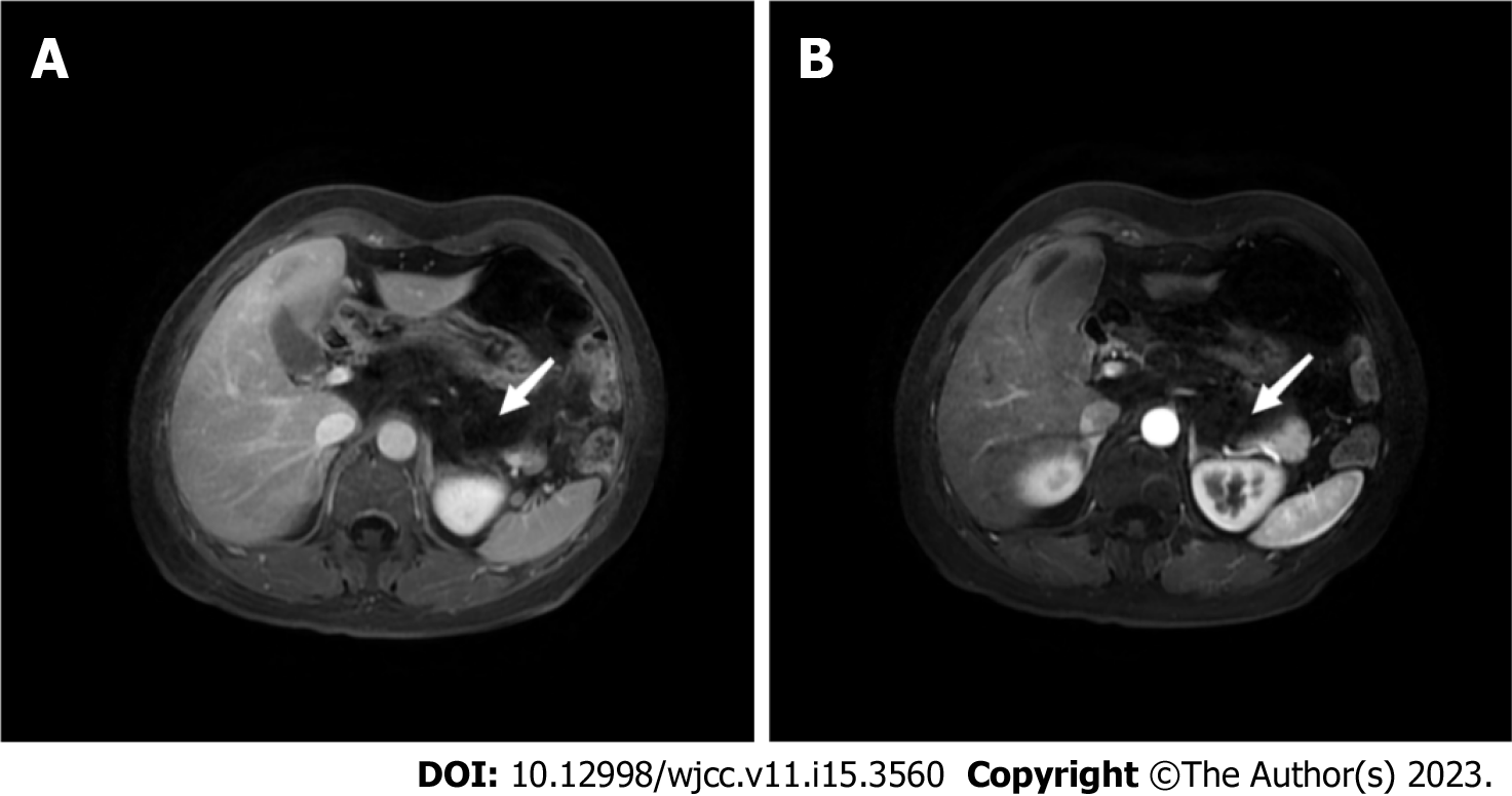
Figure 2 Magnetic resonance imaging.
A, B: The retroperitoneal layer was clear without obviously enlarged lymph nodes (arrows).
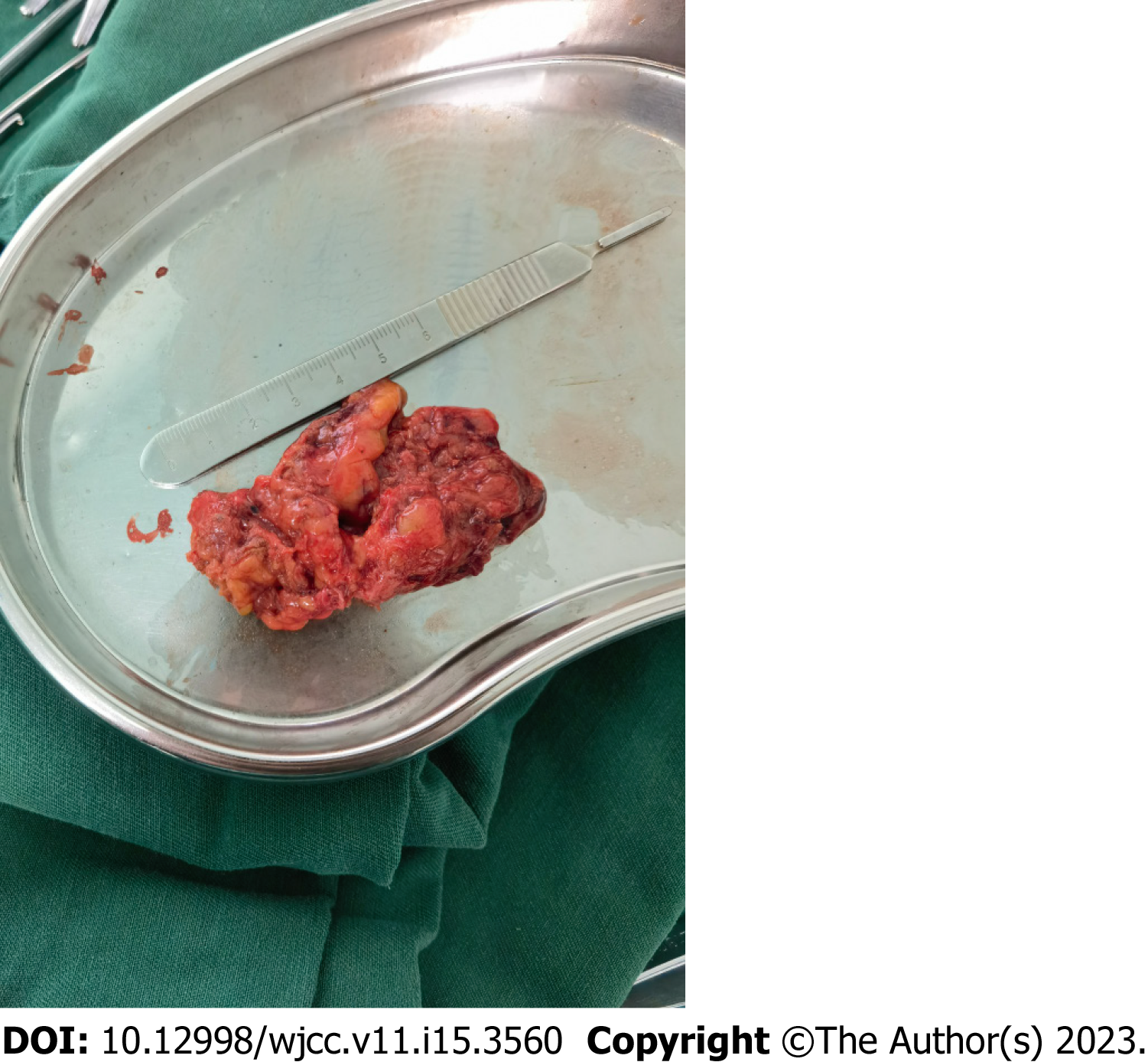
Figure 3 Resected tumor lesion.
Figure 4 Pathological examination.
A-C: Blood vessels with different lumen sizes and uneven wall thickness were seen in the tissues (arrows). A: 50×; B: 100×; C: 150×.
- Citation: Hou XF, Zhao ZX, Liu LX, Zhang H. Retroperitoneal cavernous hemangioma misdiagnosed as lymphatic cyst: A case report and review of the literature. World J Clin Cases 2023; 11(15): 3560-3570
- URL: https://www.wjgnet.com/2307-8960/full/v11/i15/3560.htm
- DOI: https://dx.doi.org/10.12998/wjcc.v11.i15.3560


