Copyright
©The Author(s) 2023.
World J Clin Cases. May 26, 2023; 11(15): 3552-3559
Published online May 26, 2023. doi: 10.12998/wjcc.v11.i15.3552
Published online May 26, 2023. doi: 10.12998/wjcc.v11.i15.3552
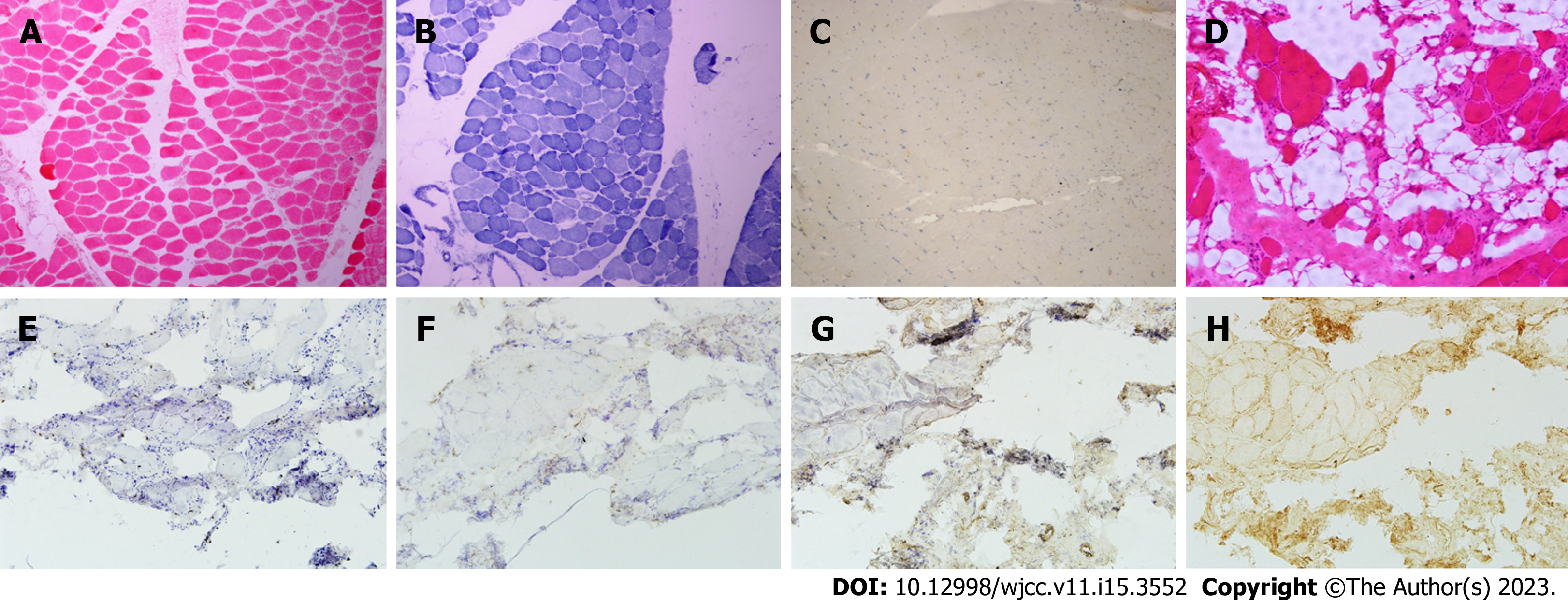
Figure 1 Muscle pathological biopsy of the patients.
A-C: Patient 1: Left anterior tibial muscle; A: Hematoxylin and eosin (HE) staining showed multiple muscle bundles. The size of muscle fibres was significantly different, most of the muscle fibres became smaller and rounder, and there were scattered necrotic and degenerative muscle fibres; B: Reduced coenzyme I staining showed some muscle fibre myofibrillar network disorders; C: Major histocompatiblility complex-1 (MHC-1) staining showed no obvious abnormalities; D-H: Patient 2: Left biceps brachii muscle; D: HE staining showed a large number of vacuoles scattered in a small group of muscle fibres. The size of muscle fibres was obviously different. Muscle fibre morphology included a large number of muscle fibres with degenerative necrosis, sections of regenerated muscle fibres scattered in with muscle hypertrophy, and obvious muscle interstitial broadening. No inflammatory cell infiltration was observed around the blood vessels; E and F: A small number of cluster of differentiation 3 and cluster of differentiation 68 positive cells were infiltrated in the endomysium; G: Membrane attack complex was positively expressed in some myofiber membranes; H: MHC-I was expressed in many myofiber membranes.
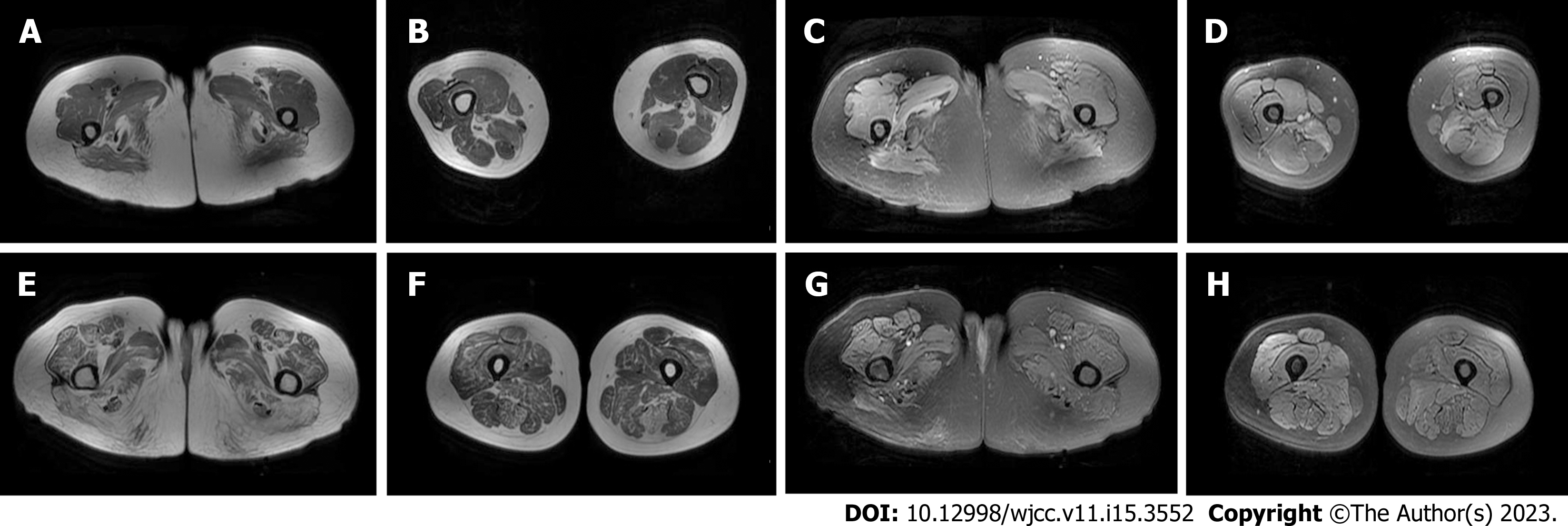
Figure 2 Magnetic resonance imaging of the thigh muscle of patients.
A-D: Patient 1; A and B: T1WI sequence showed that fat infiltration was mainly in the medial and posterior thigh muscle groups; C and D: Short time of inversion recovery (STIR) sequence showed that oedema was mainly in the posterior thigh muscle groups; E-H: Patient 2; E and F: T1WI sequence showed different degrees of fat infiltration in the muscles of the lower extremities, which was more obvious in the posterior group, and the gracilis muscle was relatively preserved; G and H: STIR sequence showed that oedema was found in the right lower limb, mainly concentrated in the anterior external and posterior thigh muscle groups.
- Citation: Chen BH, Zhu XM, Xie L, Hu HQ. Immune-mediated necrotizing myopathy: Report of two cases. World J Clin Cases 2023; 11(15): 3552-3559
- URL: https://www.wjgnet.com/2307-8960/full/v11/i15/3552.htm
- DOI: https://dx.doi.org/10.12998/wjcc.v11.i15.3552


