Copyright
©The Author(s) 2023.
World J Clin Cases. May 26, 2023; 11(15): 3533-3541
Published online May 26, 2023. doi: 10.12998/wjcc.v11.i15.3533
Published online May 26, 2023. doi: 10.12998/wjcc.v11.i15.3533
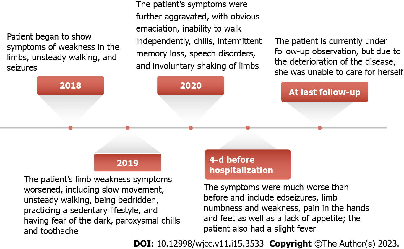
Figure 1 History of present illness.
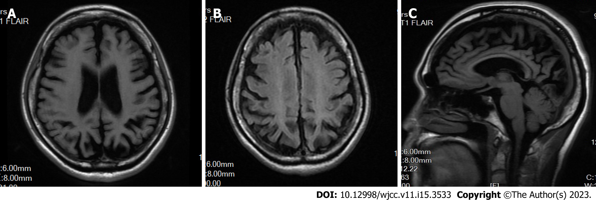
Figure 2 Brain magnetic resonance imaging showed that the patient had obvious brain atrophy.
A: T1 fluid-attenuated inversion recovery (FLAIR); B: T2 FLAIR; C: T3FLAIR.
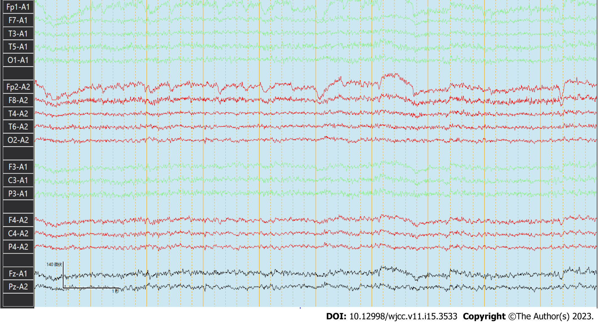
Figure 3 Electroencephalogram findings.
The basic rhythmic activity on electroencephalogram was the mid-potential 8-9c/s alpha wave and poor amplitude adjustment. Both sides were approximately equivalent. The visual response existed, and increased fast waves in both hemispheres were recorded without an obvious spike and slow wave.
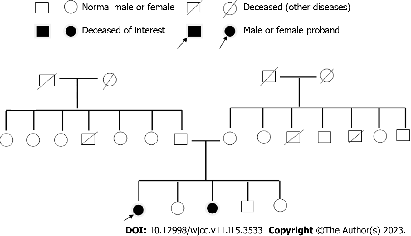
Figure 4 Pedigree of the patient.
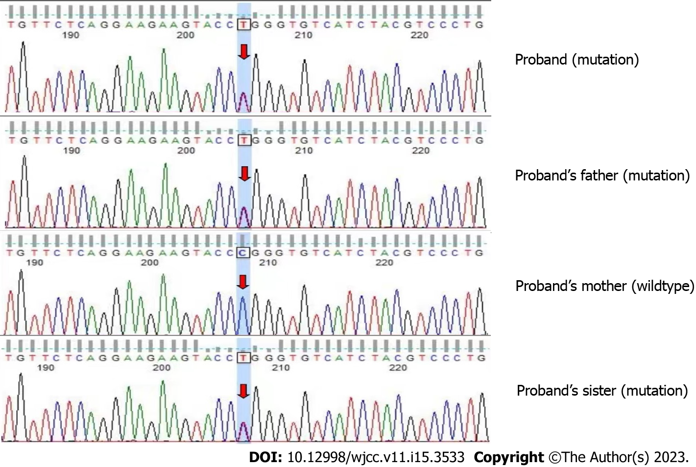
Figure 5 Whole exome sequencing revealed a mutation in the proband and the proband’s father and sister.
The mutation was CLN6 chr15:68500542 exon 7 NM_017882.3:c.872C>T(p.Pro291Leu).
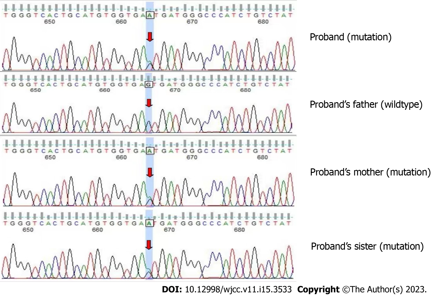
Figure 6 Whole exome sequencing revealed a mutation in the proband and the proband’s mother and sister.
The mutation was CLN6 chr15:68503596 intron 5 NM_017882.3:c.542+5G>A(p.?).
- Citation: Wang XQ, Chen CB, Zhao WJ, Fu GB, Zhai Y. Rare adult neuronal ceroid lipofuscinosis associated with CLN6 gene mutations: A case report. World J Clin Cases 2023; 11(15): 3533-3541
- URL: https://www.wjgnet.com/2307-8960/full/v11/i15/3533.htm
- DOI: https://dx.doi.org/10.12998/wjcc.v11.i15.3533


