Copyright
©The Author(s) 2023.
World J Clin Cases. May 26, 2023; 11(15): 3481-3490
Published online May 26, 2023. doi: 10.12998/wjcc.v11.i15.3481
Published online May 26, 2023. doi: 10.12998/wjcc.v11.i15.3481
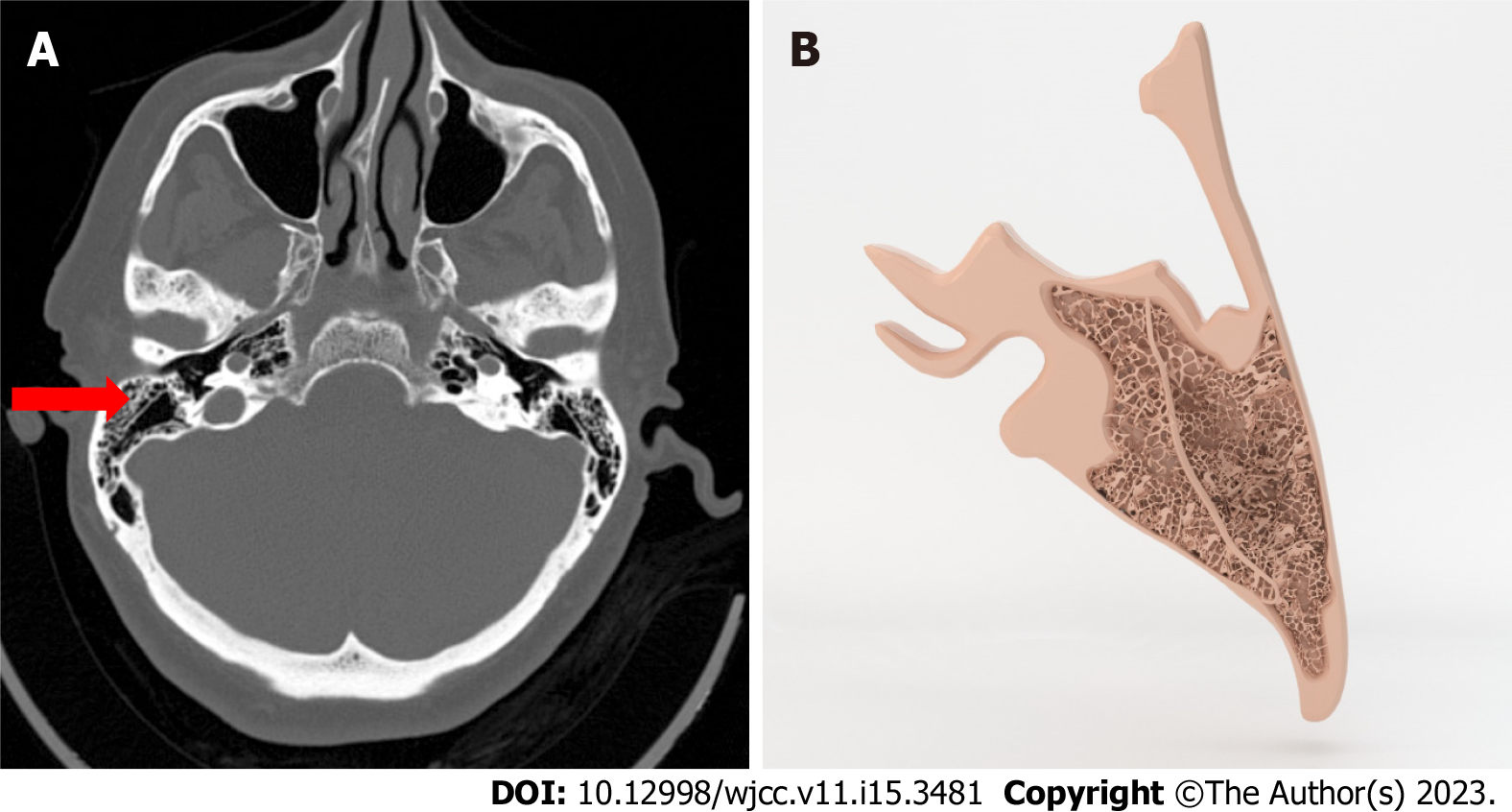
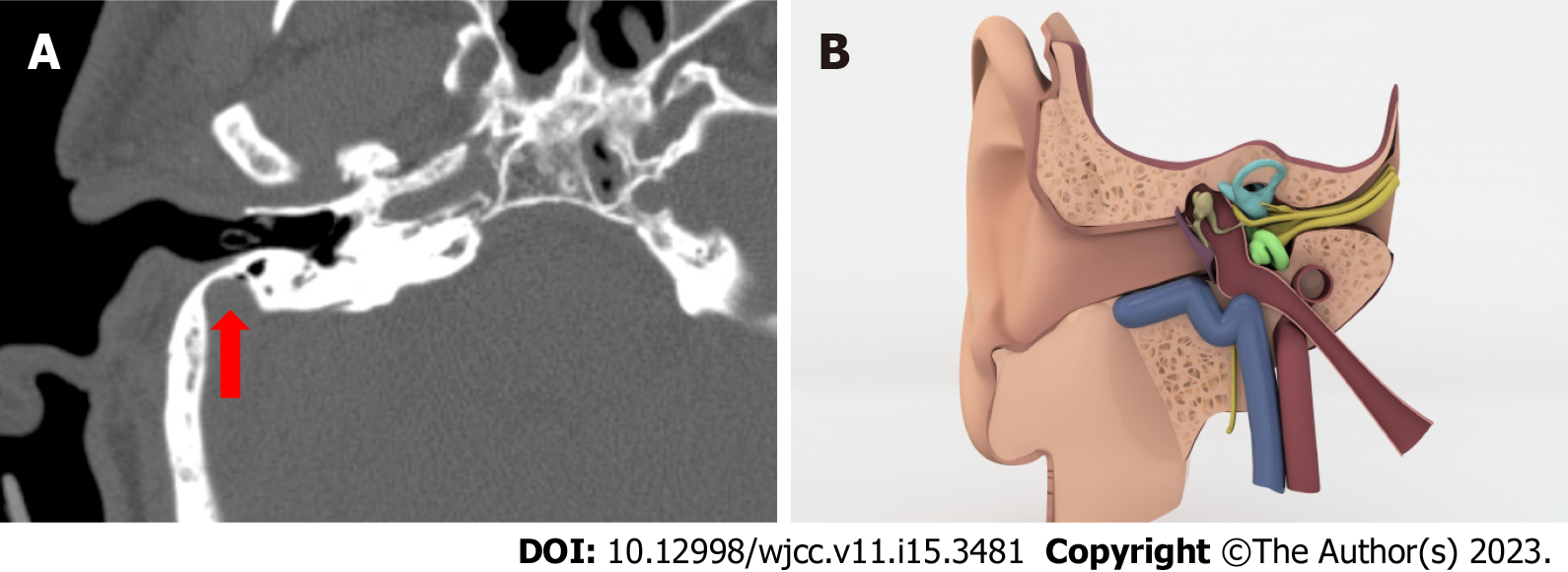
Figure 1 Koerner’s septum in axial computed tomography image and illustration.
A: Axial computed tomography image shows Koerner’s septum (KS) as a thicker bone lamina in mastoid process (red arrow); B: KS is seen in illustration.
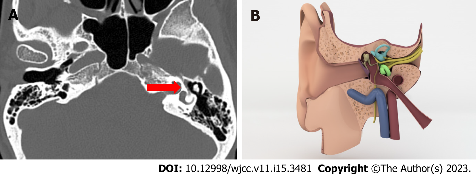
Figure 2 Labyrinth segment of facial canal without a bonny canal coverage in axial computed tomography image and illustration.
A: Axial computed tomography image shows that there is not a bonny canal coverage around the labyrinth segment of facial nerve (red arrow); B: In this illustration labyrinth segment of facial canal can be seen without a bonny canal coverage.
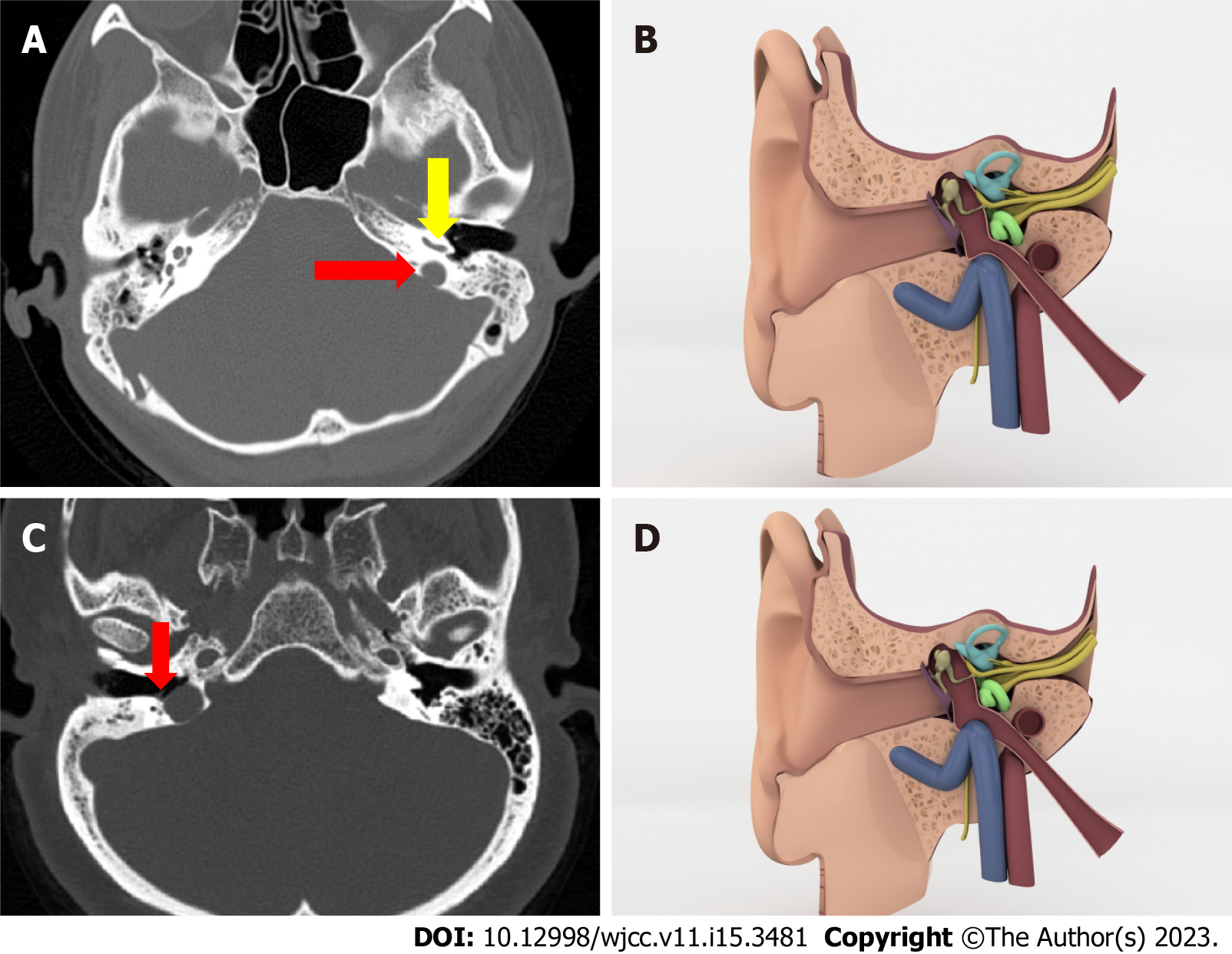
Figure 3 Axial computed tomography image and illustration.
A: Axial computed tomography image shows that the roof of the jugular bulb (red arrow) is much higher than the basal turn of cochlea (yellow arrow); B: Illustration of middle ear cavity in coronal plane show jugular bulb colored as blue is slightly higher than the basal turn of cochlea; C: Axial computed tomography image shows that there is not a bone roof above jugular bulb and it is in direct contact with middle ear cavity (red arrow), tympanic membrane; D: When comparing with B we cannot see the bonny roof of jugular bulb.
Figure 4 Jugular bulb diverticulum.
A: Axial computed tomography image shows jugular bulb diverticulum as an outpouching from tiny bulbus (red arrow); B: Illustration draw of jugular bulb diverticulum which out pouches to middle ear cavity.
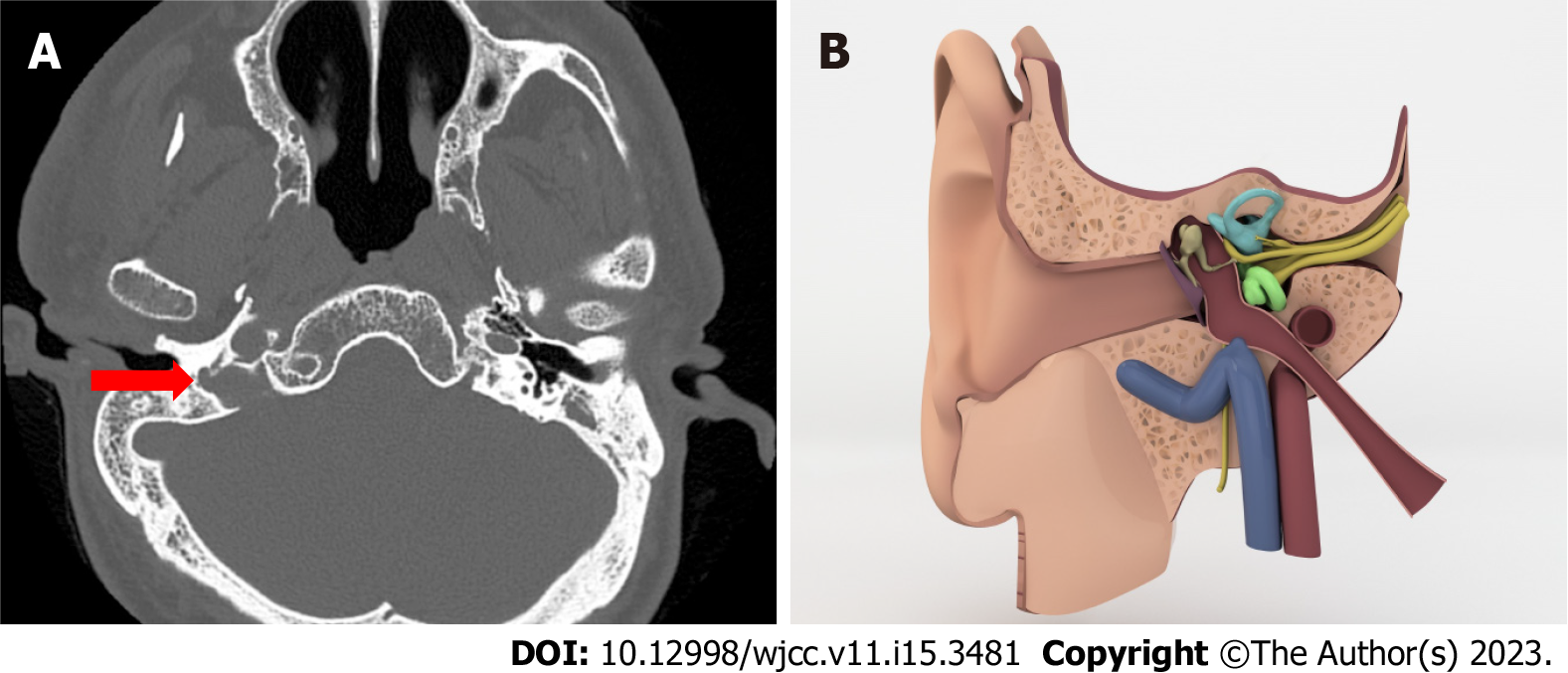
Figure 5 Anteriorly located sigmoid sinus.
A: Anteriorly located sigmoid sinus. On axial CT images shows a sigmoid sinus very close to outer ear (red arrow); B: Illustration demonstrates anteriorly located sigmoid sinus.
- Citation: Gökharman FD, Şenbil DC, Aydin S, Karavaş E, Özdemir Ö, Yalçın AG, Koşar PN. Chronic otitis media and middle ear variants: Is there relation? World J Clin Cases 2023; 11(15): 3481-3490
- URL: https://www.wjgnet.com/2307-8960/full/v11/i15/3481.htm
- DOI: https://dx.doi.org/10.12998/wjcc.v11.i15.3481


