Copyright
©The Author(s) 2023.
World J Clin Cases. May 26, 2023; 11(15): 3369-3384
Published online May 26, 2023. doi: 10.12998/wjcc.v11.i15.3369
Published online May 26, 2023. doi: 10.12998/wjcc.v11.i15.3369
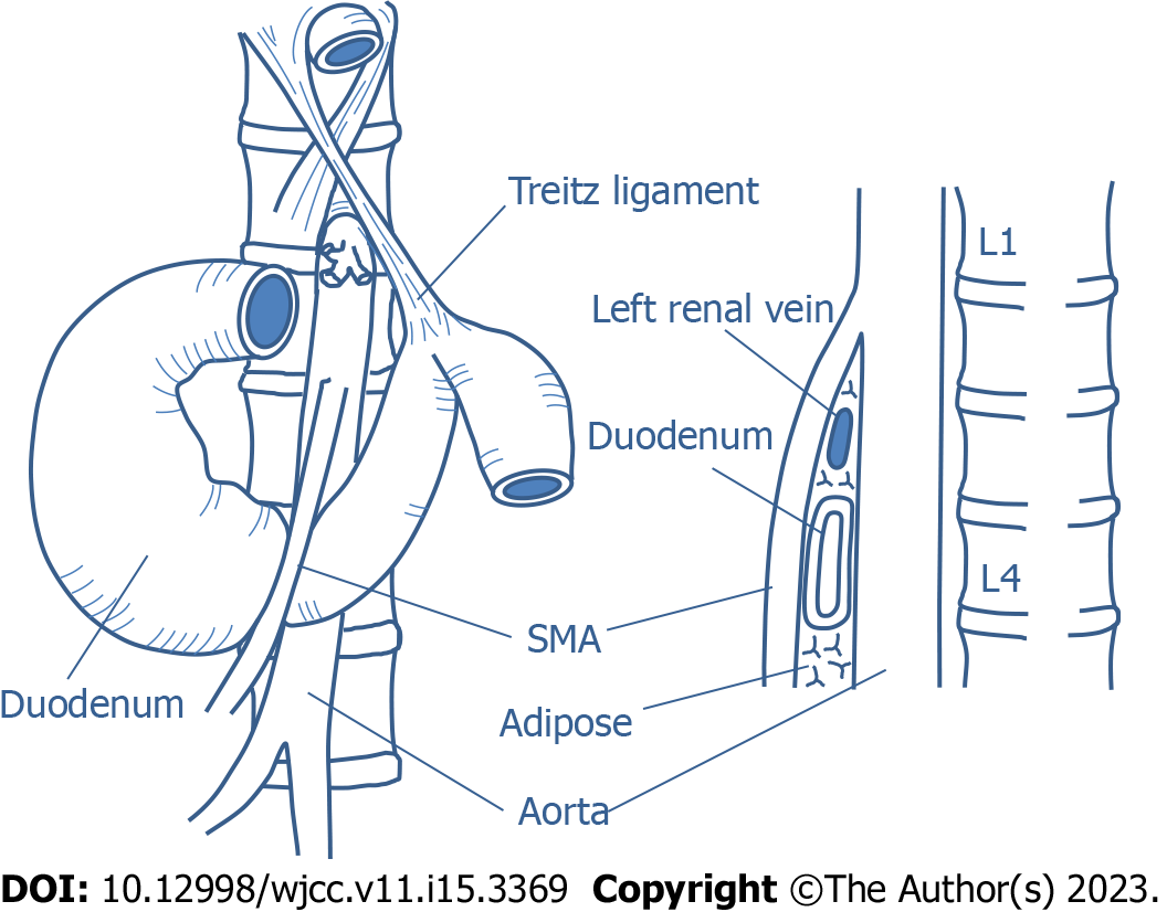
Figure 1 Anatomy related to superior mesenteric artery syndrome.
SMA: Superior mesenteric artery.
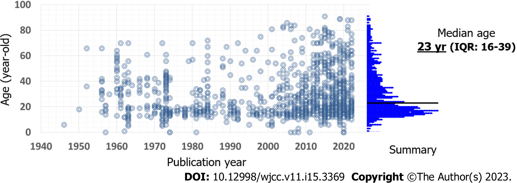
Figure 2 Published patient age.
Dot indicates an individual case. The data is based on our review. IQR: Interquartile range.
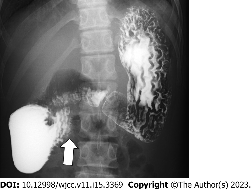
Figure 3 Upper gastrointestinal series (barium X-ray) of a 16-year-old male with superior mesenteric artery syndrome.
Arrow indicates obstructive compression of the third portion of the duodenum.
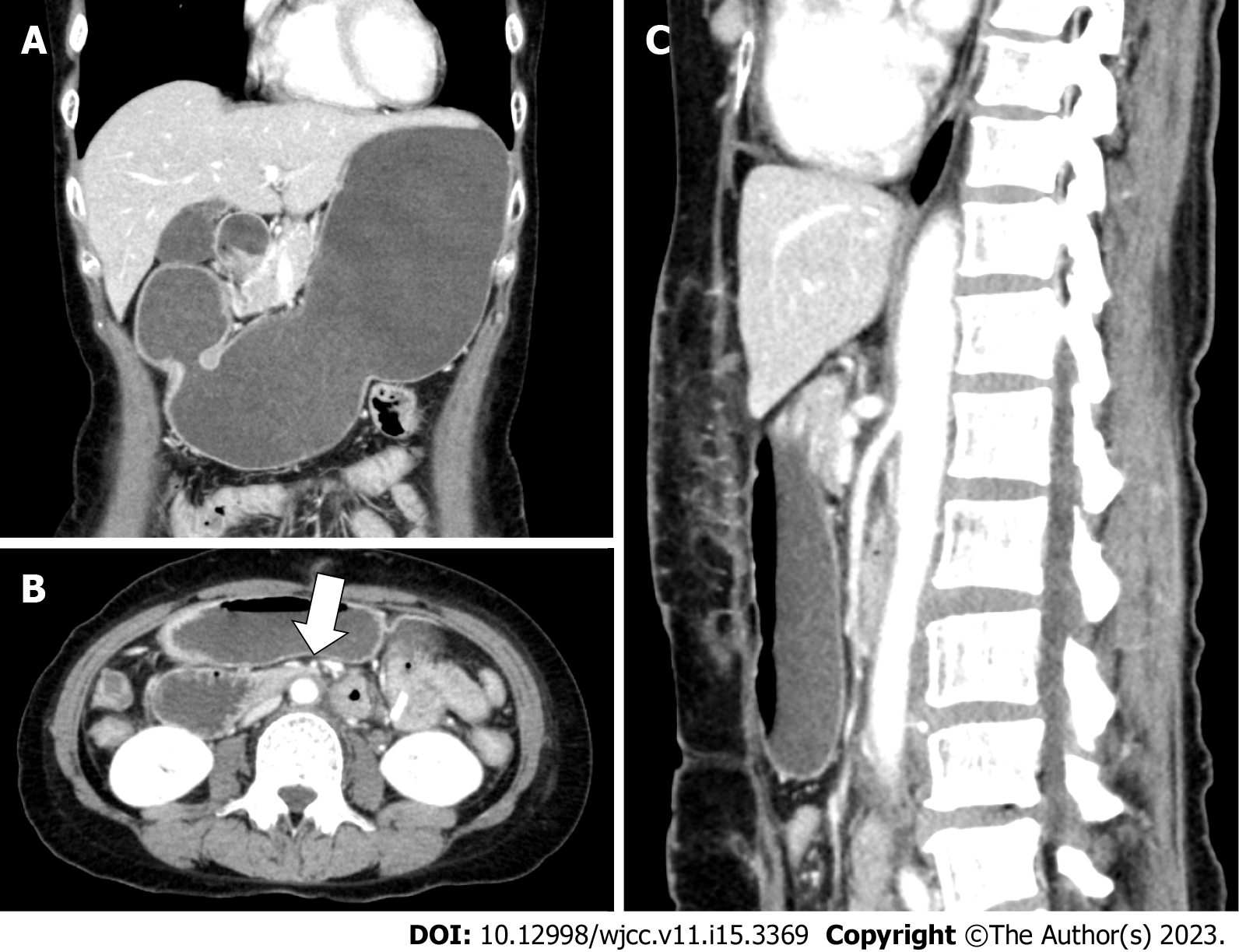
Figure 4 Enhanced computed tomography images of a 56-year-old female with superior mesenteric artery syndrome.
A: Coronal view; B: Axial view; C: Sagittal view. Computed tomography images show a markedly distended stomach and proximal duodenum by extrinsic compression between the superior mesenteric artery (arrow in panel B) and aorta.
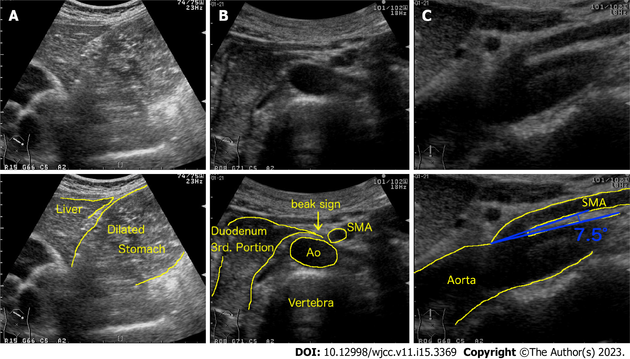
Figure 5 Abdominal ultrasonographic images of a 53-year-old female with superior mesenteric artery syndrome.
A and B: Upper abdominal ultrasonography shows a markedly dilated stomach (A) and obstruction of duodenum (B, which looks like beak, beak sign) by extrinsic compression between the superior mesenteric artery (SMA) and aorta (Ao); C: The SMA-Ao angle (7.5 degree) and distance (5 mm) are decreased. SMA: Superior mesenteric artery; Ao: Aorta.
Figure 6 Endoscopic findings of patients with superior mesenteric artery syndrome.
A and B: Esophagitis (A) and gastric ulcer (B) with retained luminal contents; C: compression area in the third portion of the duodenum.
- Citation: Oka A, Awoniyi M, Hasegawa N, Yoshida Y, Tobita H, Ishimura N, Ishihara S. Superior mesenteric artery syndrome: Diagnosis and management. World J Clin Cases 2023; 11(15): 3369-3384
- URL: https://www.wjgnet.com/2307-8960/full/v11/i15/3369.htm
- DOI: https://dx.doi.org/10.12998/wjcc.v11.i15.3369


