Copyright
©The Author(s) 2023.
World J Clin Cases. Apr 26, 2023; 11(12): 2848-2854
Published online Apr 26, 2023. doi: 10.12998/wjcc.v11.i12.2848
Published online Apr 26, 2023. doi: 10.12998/wjcc.v11.i12.2848
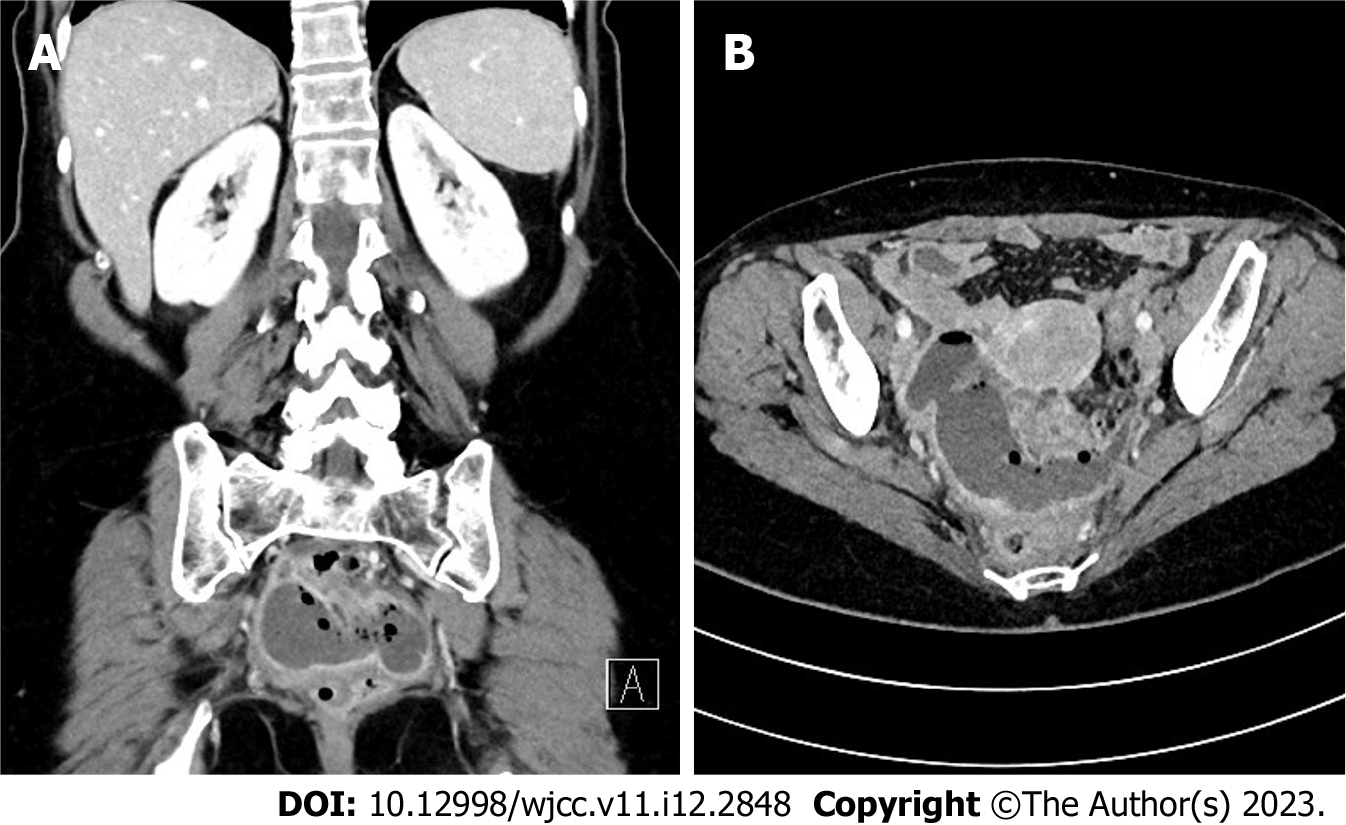
Figure 1 Abdominal computed tomography scan in axial and coronal view.
The well-encapsulated pelvic abscess measuring 8 cm × 8 cm × 5 cm is seen as a dense air-fluid collection in the proximity of the sigmoid colon. A: Axial view; B: Coronal view.
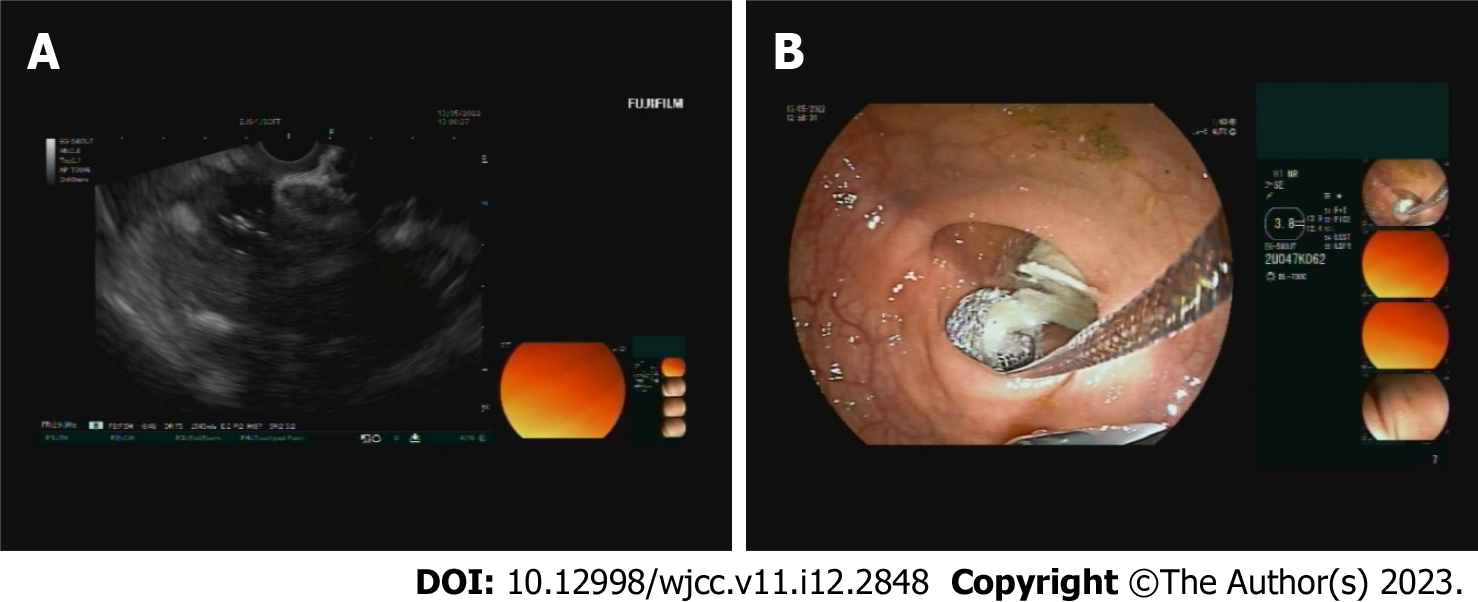
Figure 2 Endoscopic ultrasound scan during lumen-apposing metal stent insertion.
A: An Endoscopic ultrasound image of the inserted lumen-apposing metal stent (LAMS), connecting the bowel lumen and the abscess collection; B: An endoscopic view of in-situ LAMS, draining the pus from the abscess collection.
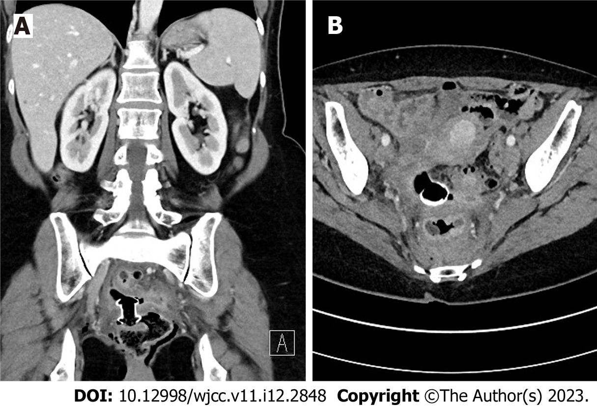
Figure 3 Follow-up abdominal computed tomography scan in axial and coronal views.
The lumen-apposing metal stent position is appropriate and lies transluminal in the sigmoid wall. Regression of the collection is seen with only some gas remaining at the site. A: Axial view; B: Coronal view.
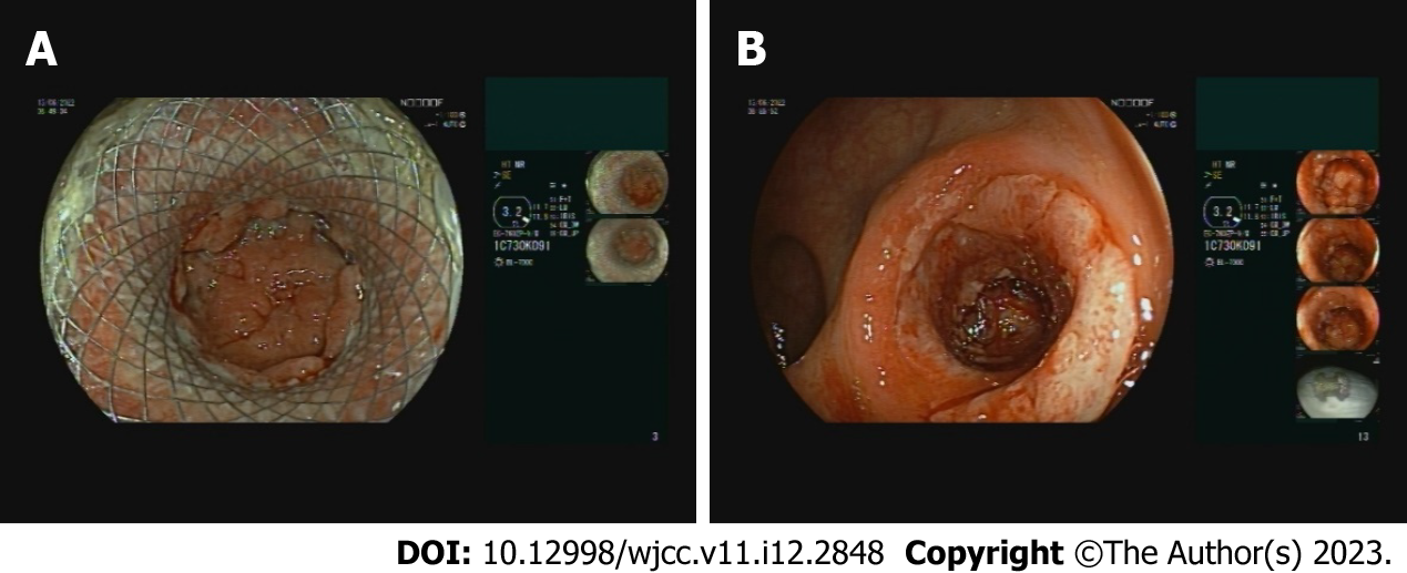
Figure 4 The findings during the removal of the lumen-apposing metal stent.
A: The lumen-apposing metal stent (LAMS) is seen in situ with regression of the abscess cavity; B: The remaining fistular canal is covered with granulation tissue after the removal of LAMS.
- Citation: Drnovšek J, Čebron Ž, Grosek J, Janež J. Endoscopic ultrasound-guided transrectal drainage of a pelvic abscess after Hinchey II sigmoid colon diverticulitis: A case report. World J Clin Cases 2023; 11(12): 2848-2854
- URL: https://www.wjgnet.com/2307-8960/full/v11/i12/2848.htm
- DOI: https://dx.doi.org/10.12998/wjcc.v11.i12.2848


