Copyright
©The Author(s) 2023.
World J Clin Cases. Apr 26, 2023; 11(12): 2788-2795
Published online Apr 26, 2023. doi: 10.12998/wjcc.v11.i12.2788
Published online Apr 26, 2023. doi: 10.12998/wjcc.v11.i12.2788
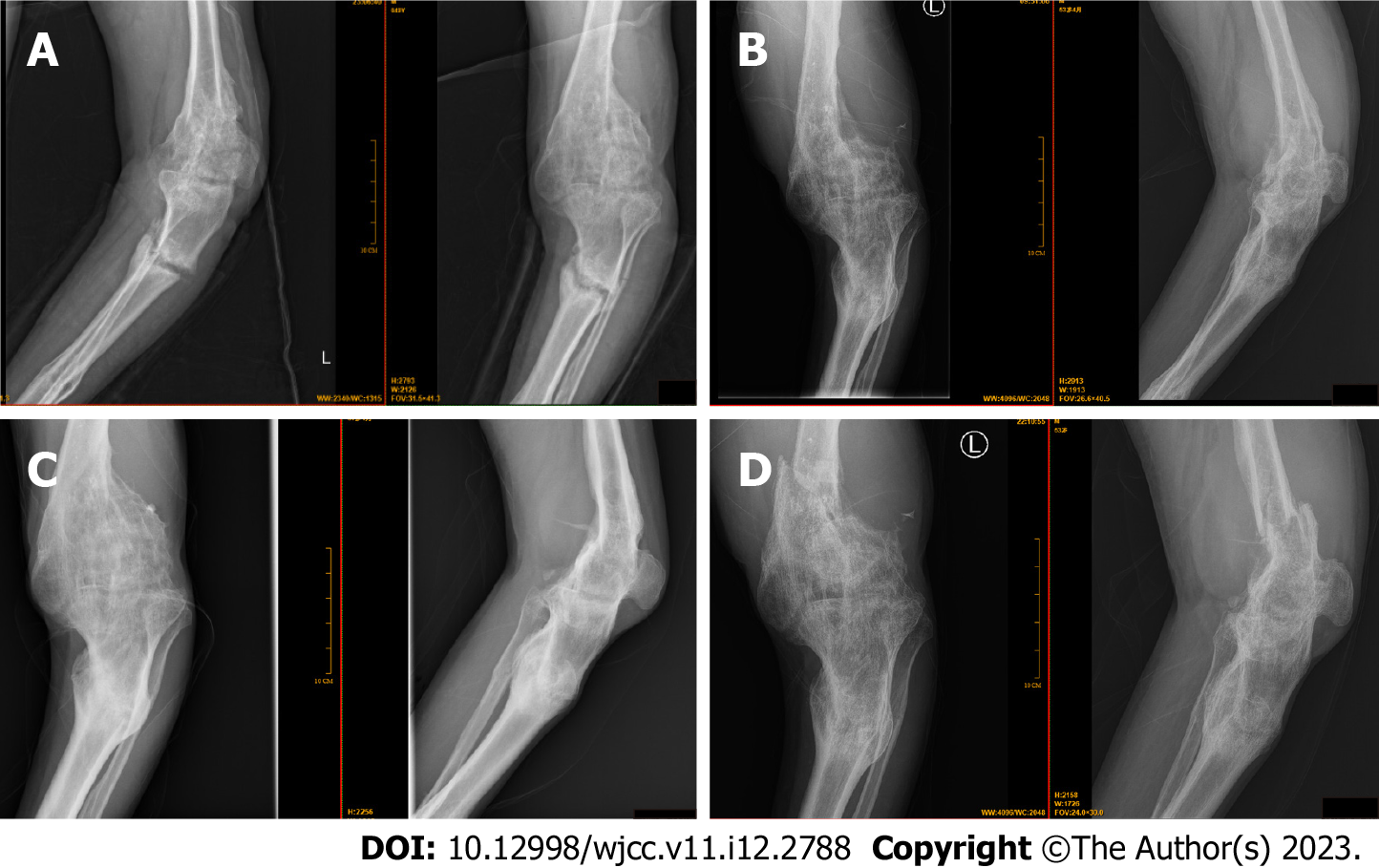
Figure 1 Typical radiographs of the patient from 2013 when he first come to our clinic.
A: Preoperative left knee anteroposterior and lateral radiographs in 2013, showed hemophiliac knee arthritis, old tibia and fibula fractures, and deformity of the distal femur fracture; B: In 2016, radiographs showed aggravated hemophiliac knee arthritis with knee fusion, deformity of the distal femur fracture, and deformity of the tibia and fibula fractures; C: On November 30, 2018, radiographs showed exacerbation of hemophiliac knee arthritis with knee fusion, deformity of the distal femur fracture, and deformity of the tibia and fibula fractures as before; D: On December 14, 2018, radiographs showed hemophiliac knee arthritis with knee fusion, deformity of the tibia and fibula fractures, and recurrent fresh fracture of the distal femur on the top of the original deformity.
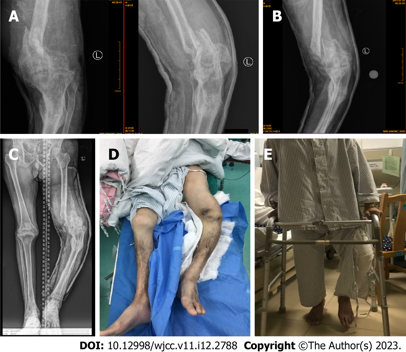
Figure 2 Preoperative radiographs of the left thigh of the patient with hemophilia.
A: On January 8, 2019, left knee anteroposterior and lateral radiographs showed hemophiliac knee arthritis with knee fusion, and recurrent fresh fracture of the distal femur was fixed with a cast; B: On January 16, 2019, left knee anteroposterior radiographs showed further displacement of the distal femoral fracture with a cast; C: Panoramic radiographs of lower limbs showed that the left thigh was nearly 8 cm shorter than the right thigh. D and E: Preoperative images of the left leg.
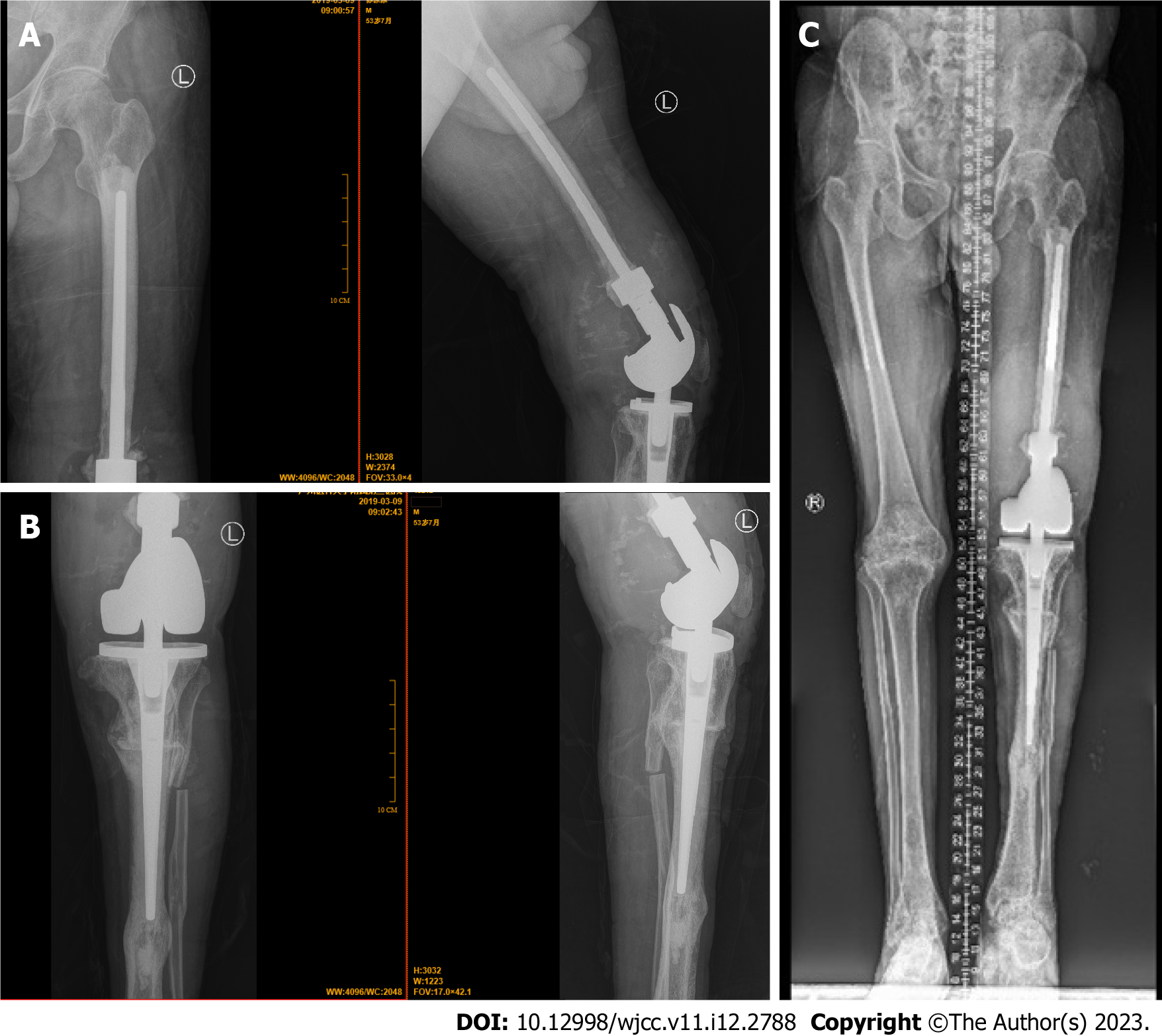
Figure 3 Postoperative radiographs of the patient with hemophilia.
A: Femur anteroposterior and lateral; B: Left knee anteroposterior and lateral; C: Panoramic view of lower limbs. The prosthesis was in the correct position with no radiographic evidence of hardware complications. The lower extremity line of strength was good, short contraction of the left lower extremity was improved, and fracture deformity of the left lower limb was corrected.
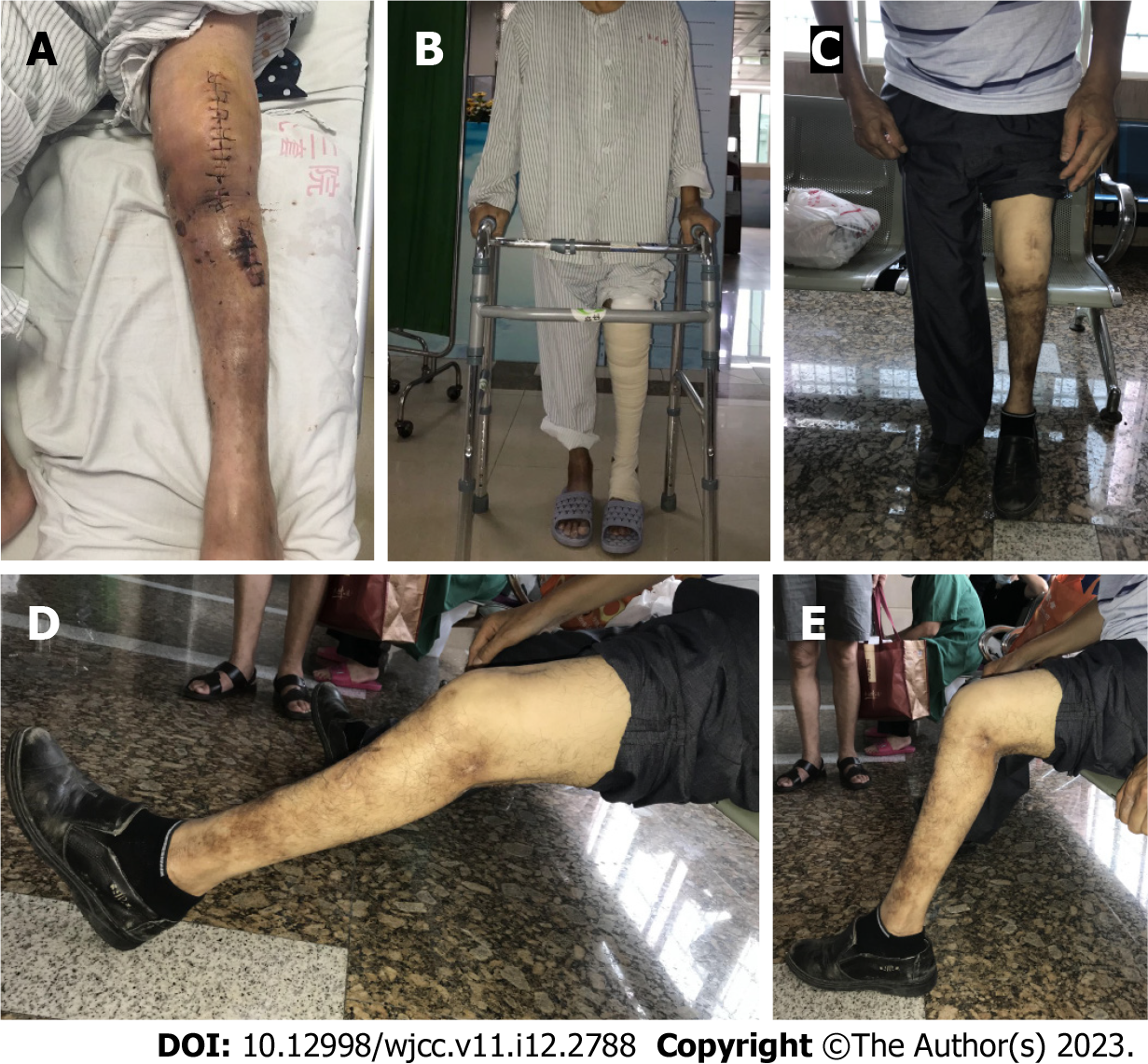
Figure 4 Post-operative and follow-up images of the patient with hemophilia.
A and B: Postoperative images of the left leg; C-E: Postoperative images demonstrated the range of motion of the left leg 16 mo after surgery. The deformity of the left leg was corrected after surgery, the length was restored, and normal function was basically restored at 16 mo after surgery.
- Citation: Yin DL, Lin JM, Li YH, Chen P, Zeng MD. Short-term outcome of total knee replacement in a patient with hemophilia: A case report and review of literature. World J Clin Cases 2023; 11(12): 2788-2795
- URL: https://www.wjgnet.com/2307-8960/full/v11/i12/2788.htm
- DOI: https://dx.doi.org/10.12998/wjcc.v11.i12.2788


