Copyright
©The Author(s) 2023.
World J Clin Cases. Jan 6, 2023; 11(1): 225-232
Published online Jan 6, 2023. doi: 10.12998/wjcc.v11.i1.225
Published online Jan 6, 2023. doi: 10.12998/wjcc.v11.i1.225
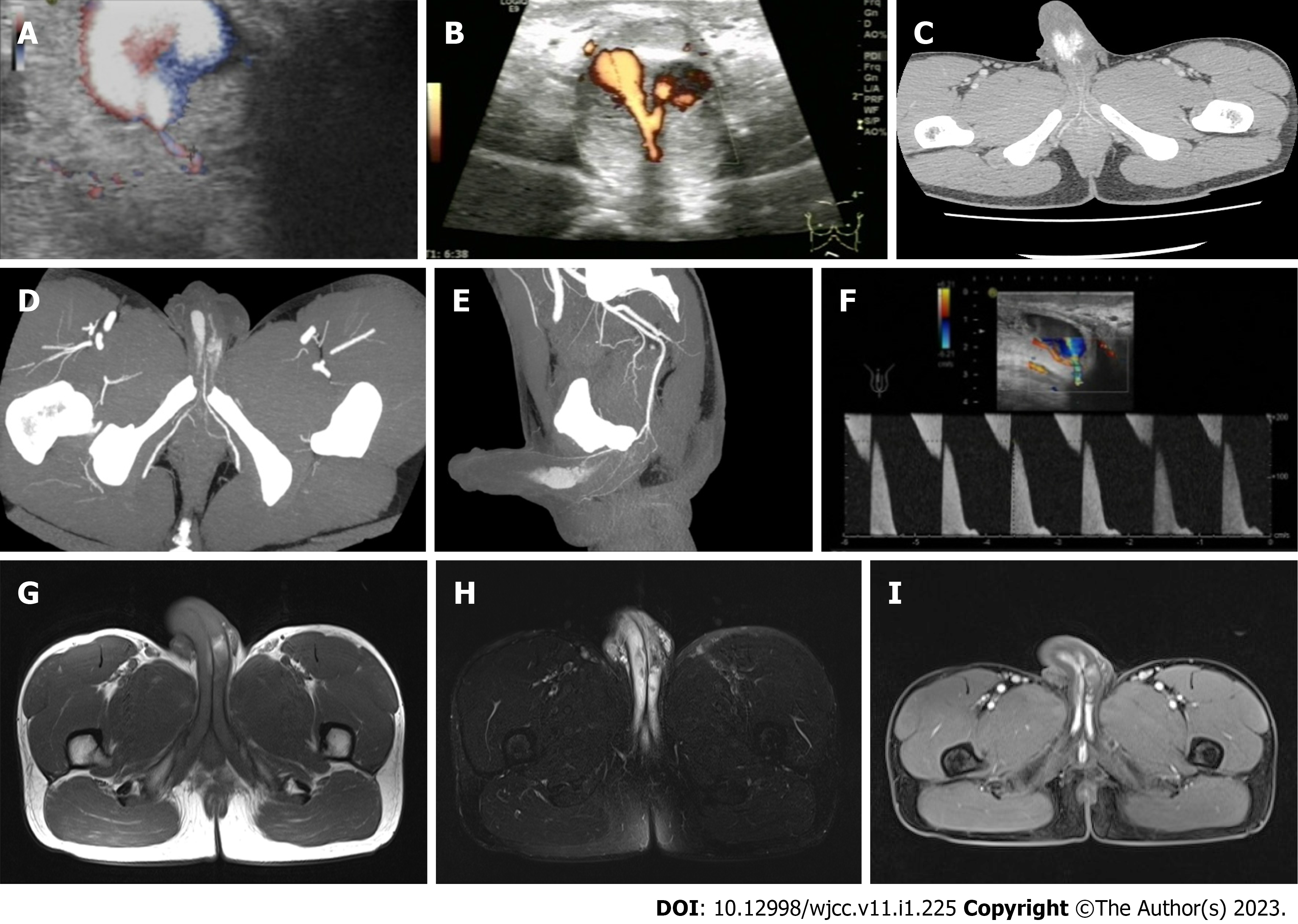
Figure 1 Color Doppler ultrasonography, computed tomography angiography, and magnetic resonance imaging of the patient.
A and B: Color Doppler ultrasonography revealed bilateral cavernous artery fistulas and bilateral pseudoaneurysms complicated by an interaneurysmal fistula (the width of the right fistula was approximately 0.6 cm); C: Contrast-enhanced computed tomography scanning (arterial phase) revealed bilateral cavernous artery pseudoaneurysms; D and E: Maximum intensity projection images revealed pelvic and penile artery anatomy; F: Arterial flow was characterized by a high flow with high resistance and irregular biphasic bidirectional flow (peak arterial flow: 162 cm/s); G: Axial T1-weighted imaging; H: Diffusion weighted imaging; I: Contrast-enhanced T1-weighted imaging (arterial phase) revealed bilateral cavernous pseudoaneurysms with cavernous septum injuries.
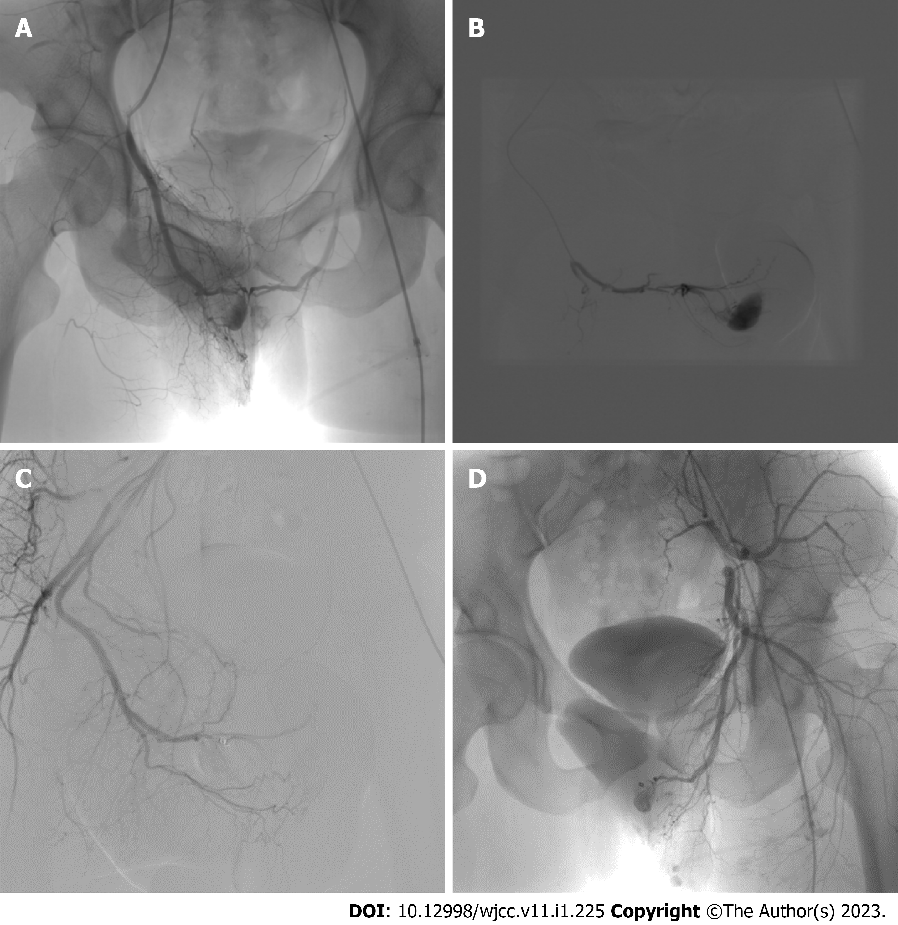
Figure 2 Digital subtraction angiography images during patient intervention.
A: Digital subtraction angiography of the patient revealed bilateral cavernous artery fistulas and pseudoaneurysms complicated by interaneurysmal fistulas; B: A microcatheter (2.7 F) was superselectively inserted into the right pudendal artery; C: Arterial embolization was performed with a microcoil (1 mm) combined with gel-foam particles (750-1200 µm) at the proximal position of the right penile dorsal artery. This led to the disappearance of the right cavernous artery fistula; D: The left cavernous artery fistula was not treated by embolization.
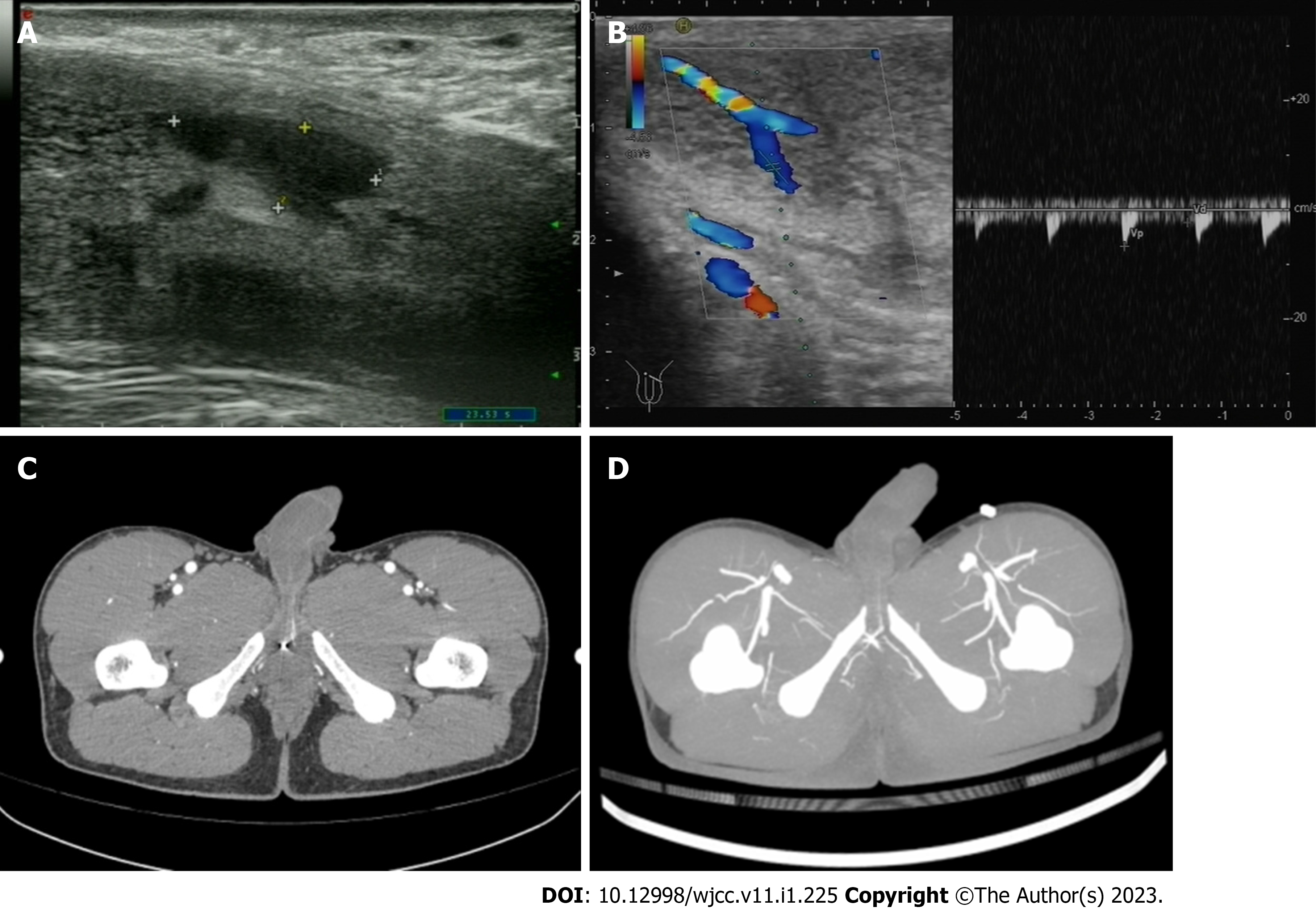
Figure 3 Color Doppler ultrasonography and computed tomography angiography of the patient 6 mo after interventional embolization.
A: Ultrasound revealed anechoic areas of the bilateral cavernous body; B: Color Doppler revealed an increased resistive index in the right dorsal penile artery (embolization side); C: Contrast-enhanced computed tomography scanning (arterial phase) revealed that the microcoil was fixed in the right penile dorsal artery; D: Maximum intensity projection images revealed that the distal part of the right penile dorsal artery had faded.
- Citation: Li G, Liu Y, Wang HY, Du FZ, Zuo ZW. High-flow priapism due to bilateral cavernous artery fistulas treated by unilateral embolization: A case report. World J Clin Cases 2023; 11(1): 225-232
- URL: https://www.wjgnet.com/2307-8960/full/v11/i1/225.htm
- DOI: https://dx.doi.org/10.12998/wjcc.v11.i1.225


