Copyright
©The Author(s) 2023.
World J Clin Cases. Jan 6, 2023; 11(1): 201-209
Published online Jan 6, 2023. doi: 10.12998/wjcc.v11.i1.201
Published online Jan 6, 2023. doi: 10.12998/wjcc.v11.i1.201
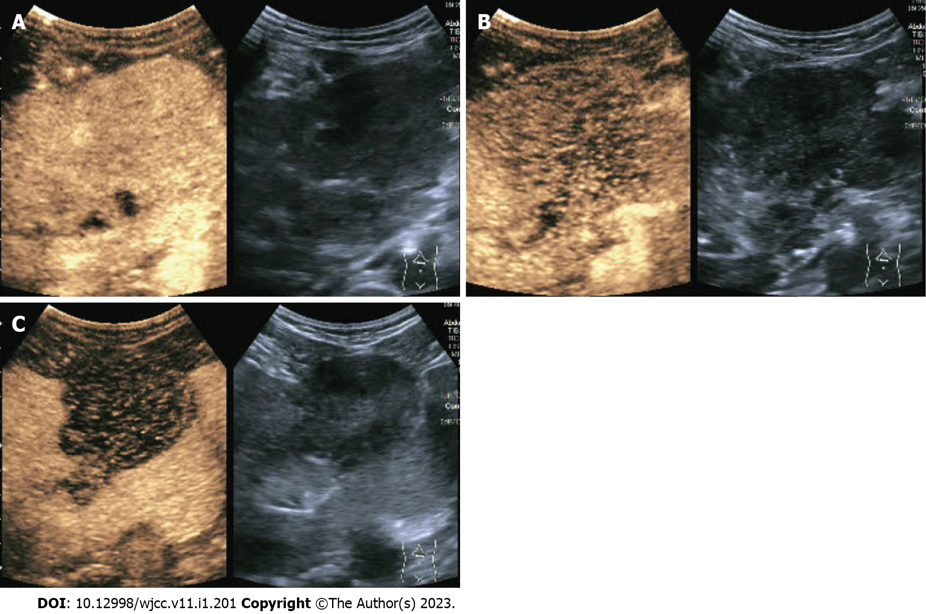
Figure 1 Abdominal contrast-enhanced ultrasound.
A: Hepatic arterial phase; B: Hepatic portal vein phase; C: Hepatic delay phase.
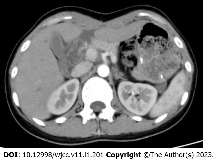
Figure 2 Contrast-enhanced abdominal computerized tomography.
A: Hepatic arterial phase; B: Hepatic portal phase; C: Hepatic venous phase.
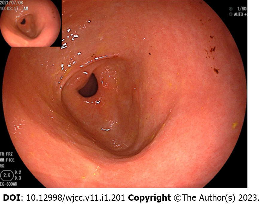
Figure 3 Esophagogastroduodenoscopy revealed that the gastric antrum mucosa was intact and smooth.
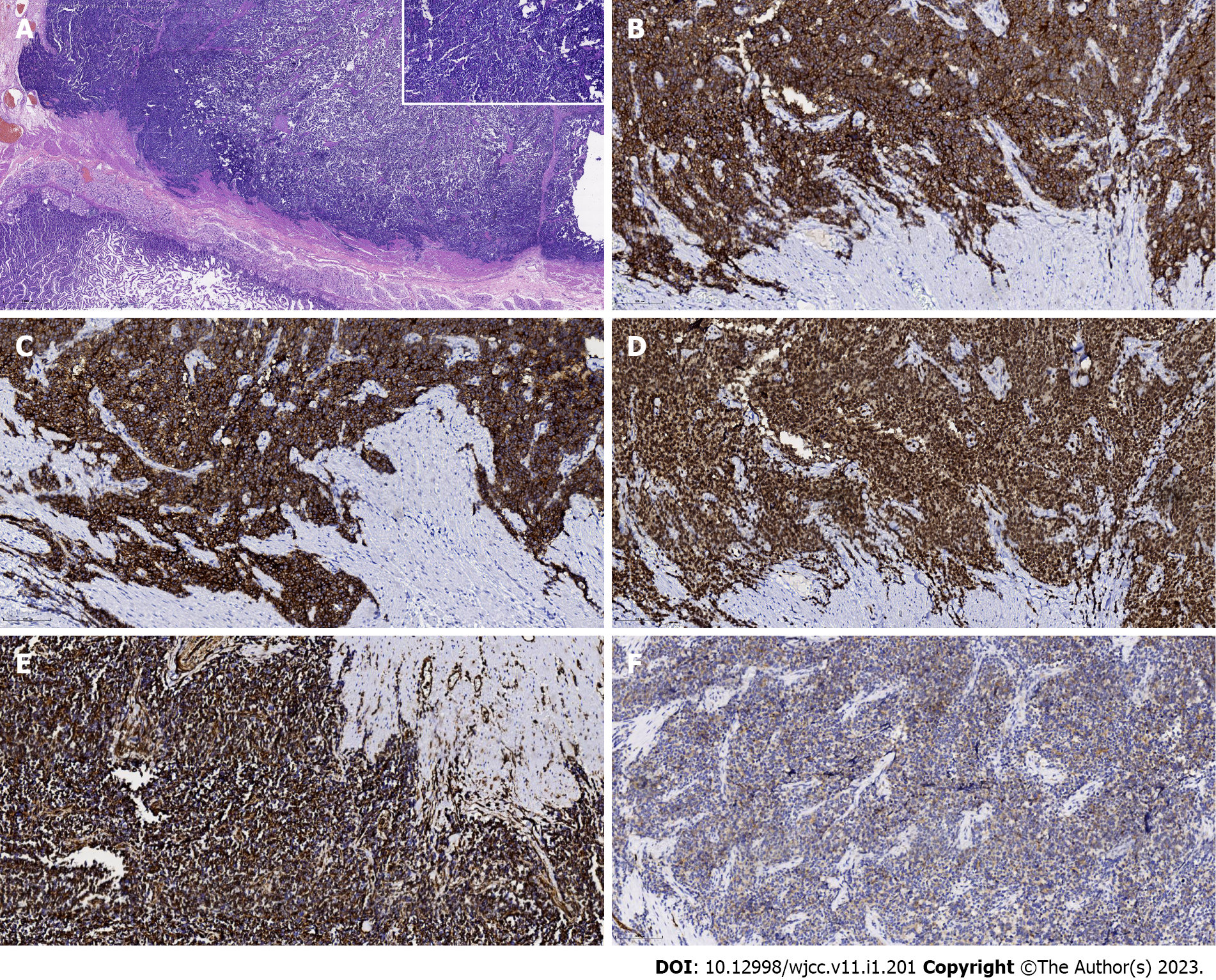
Figure 4 Postoperative pathological examination.
A: Hematoxylin and eosin staining. The tumor was identified to originate from the serous layer of the stomach and involve the muscularis externa of the stomach (20 ×). Upper right inset indicates small round cells with different sizes (original magnification, 200 ×); B-F: CD99-positive, vimentin-positive, FLI-1-positive, NSE-positive, amd SYN-positive cells (immunohistochemistry staining, 200 ×).
Figure 5 Contrast-enhanced abdominal computerized tomography 11 mo postoperatively.
- Citation: Shu Q, Luo JN, Liu XL, Jing M, Mou TG, Xie F. Extraskeletal Ewing sarcoma of the stomach: A rare case report. World J Clin Cases 2023; 11(1): 201-209
- URL: https://www.wjgnet.com/2307-8960/full/v11/i1/201.htm
- DOI: https://dx.doi.org/10.12998/wjcc.v11.i1.201


