Copyright
©The Author(s) 2023.
World J Clin Cases. Jan 6, 2023; 11(1): 193-200
Published online Jan 6, 2023. doi: 10.12998/wjcc.v11.i1.193
Published online Jan 6, 2023. doi: 10.12998/wjcc.v11.i1.193
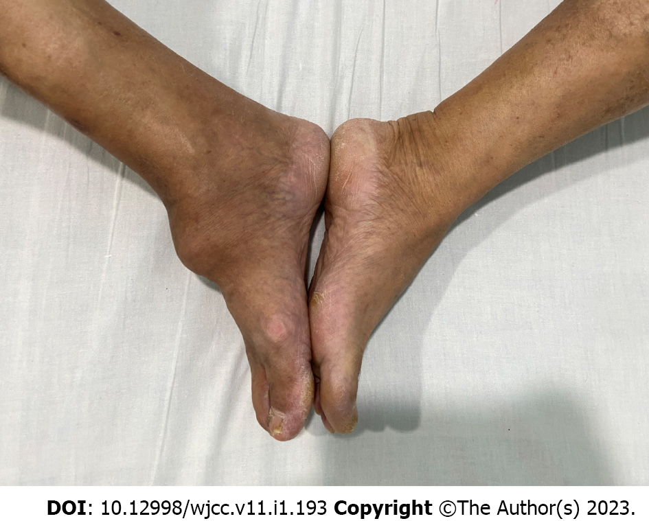
Figure 1 The appearance of heterotopic ossification in this patient.
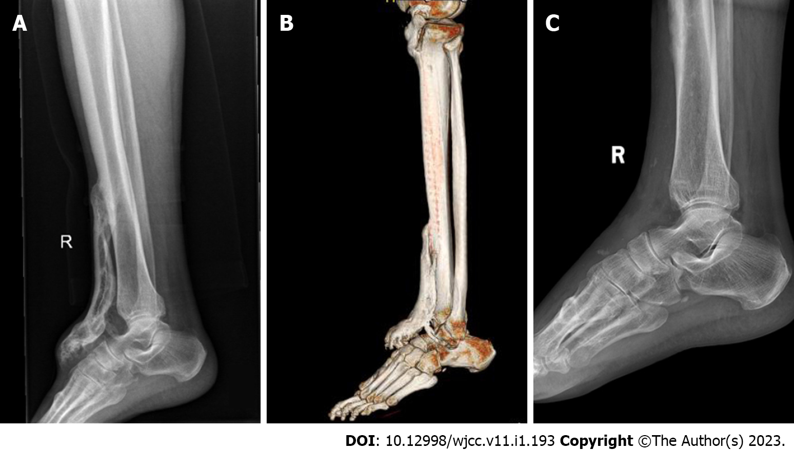
Figure 2 Radiology findings in this patient.
A: X-ray showed that a bone mass was seen in front of the right tibia; B: Computed tomography showed multiple patchy bone masses with uneven bone density in front of the upper segment of the right tibia and the dorsum of the right foot. The edge of the right ankle joint was osteosclerotic with adequate joint space; C: X-ray at follow-up showed that there was no recurrence of heterotopic ossification.
Figure 3
Biopsy of the bone mass showed ossification in the muscle tissue in some areas, which was consistent with the changes of myositis ossificans when combined with the clinical and imaging findings.
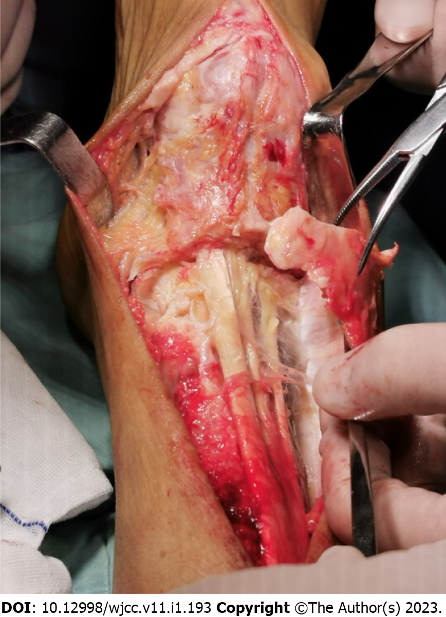
Figure 4
The extensor hallucislongus tendon formed a tunnel in the heterotopic ossification.
- Citation: Xu Z, Rao ZZ, Tang ZW, Song ZQ, Zeng M, Gong HL, Wen J. Post-traumatic heterotopic ossification in front of the ankle joint for 23 years: A case report and review of literature. World J Clin Cases 2023; 11(1): 193-200
- URL: https://www.wjgnet.com/2307-8960/full/v11/i1/193.htm
- DOI: https://dx.doi.org/10.12998/wjcc.v11.i1.193


